PET/CT > Artifacts & Pitfalls > Motion
Motion
![]()
Because the PET emission images are acquired after the CT images, any change in patient position between the two acquisitions can lead to misregistration of the PET data with the CT data. This motion will result in artifacts in the attenuation correction process. For example, the computer will not be able to localize the true body edge for attenuation correction of the portion of the PET data which does not overlie the body edge on CT data due to motion. Misregistration can also lead to inaccuracies of anatomic localization of FDG activity. Misregistration most commonly occurs in the head and limbs where movement is most likely to occur. In any case where misregistration of data due to patient motion poses a diagnostic dilemma, the study should be repeated with careful pre-examination coaching and monitoring of the patient for motion during the exam.
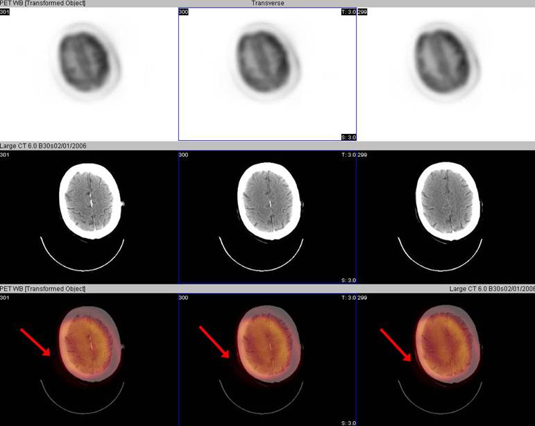
68 year old male with history of right helical (ear) melanoma. PET/CT ordered for restaging.
Patient was positioned straight for the CT acquisition, but then tilted his head rightward for the PET acquisition. Significant motion of the head between the acquisitions results in misregistration and artifact. Detailed evaluation of the head and neck is limited. The patient remained still on a repeat examination which demonstrated no abnormal FDG activity to suggest recurrent of metastatic melanoma.
Misregistration of PET and CT data can occur at the diaphragm due to respiratory motion and associated movement of adjacent organs above and below the diaphragm. Because the CT scan is acquired during breath-hold with a moderate inspiration and the PET scan is acquired over several minutes during tidal respiration, the diaphragm and structures immediately adjacent to it are often slightly misregistered. This misalignment can cause attenuation correction artifacts and anatomic localization artifacts. The liver dome and superior aspect of the spleen appear to be "floating" above the diaphragm. Hypermetabolic foci in the dome of the liver can appear to be located in the lung base on attenuation corrected images due to diaphragmatic motion related misregistration. Also, nodules in the lung bases which are not metabolically hyperactive can be located in areas where activity in the liver dome or superior aspect of the spleen is superimposed due to diaphragmatic motion related misregistration. Therefore, it is very important to always evaluate both the non-attenuation-corrected and attenuation-corrected data sets in conjunction with the CT data to correctly localize abnormal foci of activity or exclude artifactually apparent foci of abnormal activity. However, diaphragmatic motion-related artifacts can sometimes result in equivocal findings which require other methods of evaluation.
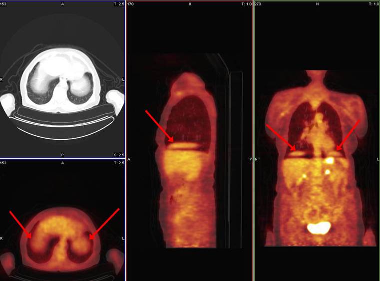
81 year old female with history of gastrointestinal stromal tumor. Solitary pulmonary nodule seen on chest CT. PET/CT ordered for evaluation.
There was no abnormal activity within the pulmonary nodule to suggest pulmonary malignancy. Diaphragmatic motion with the so-called "floating liver and spleen" is demonstrated.
![]()
![]()