ICU Chest Films > Air in the Chest > Pneumothorax > Anteromedial Pneumothorax
Anteromedial Pneumothorax
![]()
Anteromedial pneumothoraces are differentiated into those which are superior or inferior to the pulmonary hilum. A superior anteromedial pneumothorax may result in visualization of the superior vena cava or azygos vein on the right. An inferior anteromedial pneumothorax may be evidenced by delineation of the heart border and a lucent cardiophrenic sulcus. This is the key sign of a pneumothorax as this is the highest point in the supine patient, where the air will accumulate first.
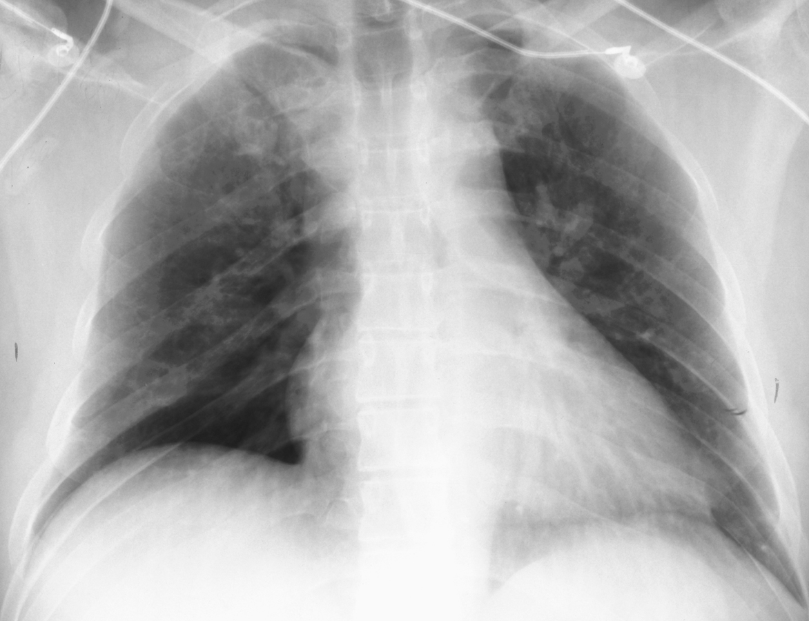
Notice the increased lucency of the cardiophrenic sulci in this patient
with bilateral
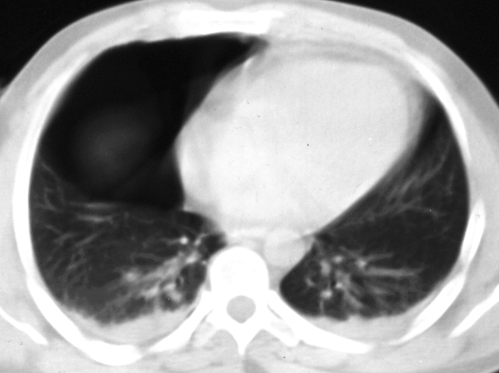
inferior anteromedial pneumothoraces. A CT scan confirms the diagnosis.
![]()
![]()