Chest Radiology > Pathology > Pulmonary Edema > CHF
Congestive
Heart Failure
![]()
Congestive heart failure (CHF) is one of the most common abnormalities evaluated by CXR. CHF occurs when the heart fails to maintain adequate forward flow. CHF may progress to pulmonary venous hypertension and pulmonary edema with leakage of fluid into the interstitium, alveoli and pleural space.
The earliest CXR finding of CHF is cardiomegaly, detected as an increased cardiothoracic ratio (>50%). In the pulmonary vasculature of the normal chest, the lower zone pulmonary veins are larger than the upper zone veins due to gravity. In a patient with CHF, the pulmonary capillary wedge pressure rises to the 12-18 mmHg range and the upper zone veins dilate and are equal in size or larger, termed cephalization. With increasing PCWP, (18-24 mm. Hg.), interstitial edema occurs with the appearance of Kerley lines. Increased PCWP above this level is alveolar edema, often in a classic perihilar bat wing pattern of density. Pleural effusions also often occur.
CXR is important in evaluating patients with CHF for development of pulmonary edema and evaluating response to therapy as well.
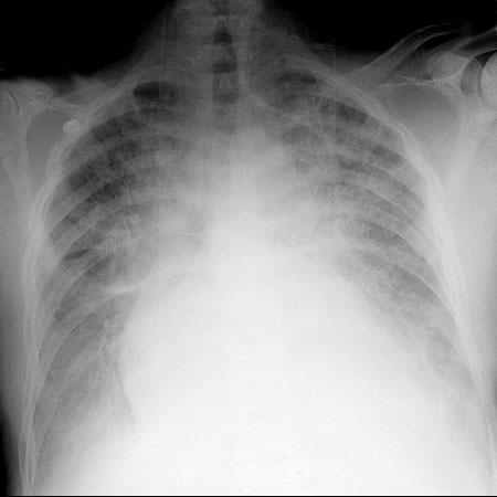
This is a typical chest x-ray of a patient in severe CHF.
Note the cardiomegaly, alveolar edema, and haziness of vascular margins.
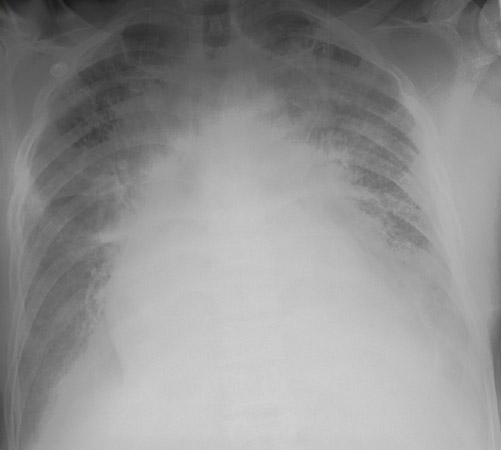
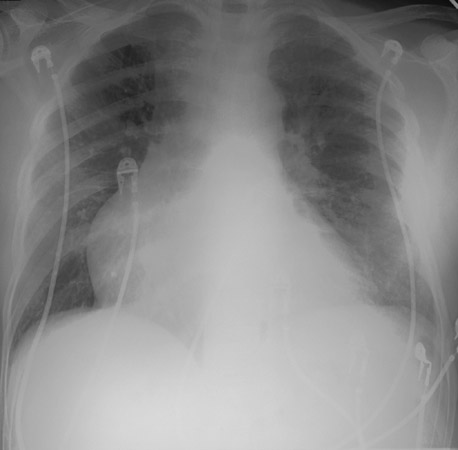
The left image demonstrates a patient with a severe pulmonary edema as a result of CHF. The right image is the same patient after significant resolution.
![]()
![]()