Cardiac MRI > Pathology > Cardiac Masses > Cardiac Thrombi
Cardiac Thrombi
![]()
Thrombi are usually formed in the heart due to atrial fibrillation or left ventricular dysfunction. Cardiac thrombi are at risk of embolizing. Cardiac MRI can diagnose both ventricular and atrial thrombi, including atrial appendage lesions.
Thrombi can often be characterized with gradient echo imaging, where they demonstrate low signal intensity relative to myocardium due to the paramagnetic effects from deoxyhemoglobin and methemoglobin which decrease the signal. An exception is fresh thrombus which can demonstrate high signal intensity. On T2-weighted spin echo images, tumors typically demonstate high signal relative to myocardium, unless they contain hemorrage (i.e. myxoma).
The primary means to differentiate thrombi from tumor is with the use of gadolinium contrast agents. Thrombi do not take up contrast agent. (Some chronic thrombi that have become organized and have undergone significant fibrosis may demonstrate some degree of enhancement, but this is a small minority of cases.)
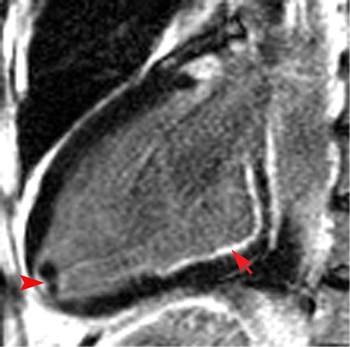
This delayed enhanced IR image shows two small thombi at the apex of the left ventricle adjacent to an apical infarct (arrowhead). They do not take up any contrast agent, which is typical of intracardiac thrombi. Also note the basal inferior wall subendocardial DHE consistent with subendocardial infarction (arrow).
![]()
![]()