Imaging of the Cervical Spine > Fractures > Jefferson Fracture (cont.)
Jefferson Fracture
![]()
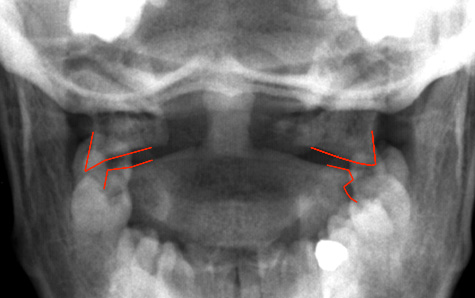
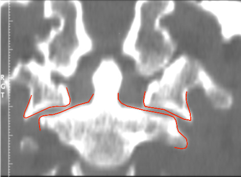
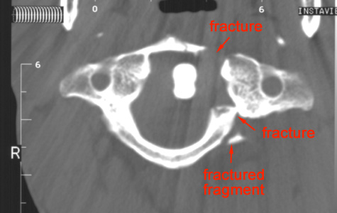
Here is an example of Jefferson fracture. The first image is the odontoid view, which illustrates the lateral displacement of C1. The second image is a coronal reconstruction from a CT, which confirms the findings from the odontoid view. The last image is a CT axial view, which clearly shows the location of the fractures of C1.


