Imaging of the Cervical Spine > Quiz Answers


Quiz Answers

alignment Question 1: The indications for getting a cervical spine series include all of the following except:
Patient is intoxicated
Patient reports midline neck pain and tenderness on palpation.
Patient has neurologic signs and symptoms.
Trauma
Explanation: Not all trauma patients with a significant injury need c-spine films. Criteria for excluding cervical spine fractures on a clinical basis are: no neck pain, no neck tenderness on palpation, having full, painless, active range of motion of c-spine, no history of loss of consciousness, no mental status change, no neurologic deficit from neck injury, and no distracting symptoms


Question 2: Visualization of C7-T1 can often be enhanced by:
Swimmer's view.
Flexion of neck.
Extension of neck.
Odontoid view.
Explanation: Visualization of C7-T1 may be limited by the amount of soft tissue in the shoulder region and can be enhanced by either applying traction on arms if no arm injury is present, or, swimmer's view (taken with one arm extended over the head). Flexion and extension views should not be obtained until the lateral view is evaluated.


Question 3: Which one of the following line is not part of the parallel lines alignment evaluation:
Anterior vertebral line.
Posterior vertebral line.
Spinolaminar line.
Facet line.
Posterior spinous line.
Explanation: see the information on alignment.


Question 4: After seeing this lateral view, the radiologist obtains a CT. This is a:

Hangman's fracture.
Jefferson fracture.
Odontoid fracture.
Teardrop fracture.
Explanation: Jefferson fracture is compression fracture of the bony ring of vertebra C1, characterized by lateral masses splitting and transverse ligament tear. The mechanism of injury is axial blow to the vertex of the head (e.g. diving injury).


Question 5: This 15 year old boy dove into a shallow pool head first and complained of neck pain. The lateral view shows a:

Hangman's fracture.
Jefferson fracture.
Odontoid fracture.
Teardrop fracture.
Explanation: Teardrop fracture usually results from hyperflexion and compression (e.g. from diving into shallow water). On lateral view, one can usually see:
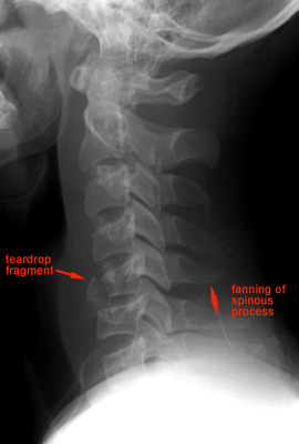
1. Prevertebral swelling associated with anterior longitudinal ligament tear.
2. Teardrop fragment from anterior vertebral body avulsion fracture.
3. Posterior vertebral body subluxation into the spinal canal.
4. Spinal cord compression from vertebral body displacement.
5. Fracture of the spinous process.


Question 6: This patient arrived at the ED after a MVA. This lateral view reveals a:

Hangman's fracture
Jefferson fracture.
Odontoid fracture.
Teardrop fracture.
Explanation: Hangman's fracture usually results from hyperextension of neck secondary from hitting head on dashboard in a MVA. Lateral view shows:
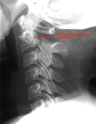
1. Prevertebral soft tissue swelling.
2. Avulsion of anterior inferior corner of C2 associated with rupture of the anterior longitudinal ligament.
3. Anterior dislocation of the C2 vertebral body.
4. Bilateral C2 pars interarticularis fractures.


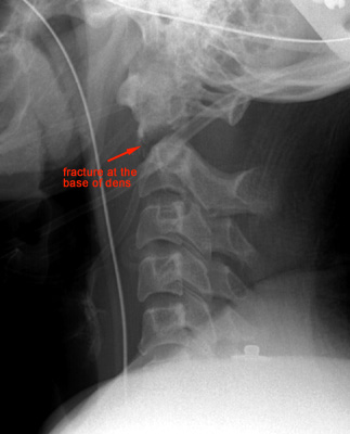
Question 7: This lateral view shows a:

Hangman's fracture.
Jefferson fracture.
Odontoid fracture.
Teardrop fracture.
Explanation: Type II Odontoid fracture is fracture at the base on the dens. The lucent fracture line is best seen on the lateral view.


Question 8: This lateral view shows a:

Hangman's fracture
Odontoid fracture.
Bilateral facet dislocation.
Unilateral facet dislocation.
Explanation: Bilateral facet dislocation is complete anterior dislocation of the vertebral body resulting from extreme hyperflexion injury. It associates with a very high risk of cord damage. Lateral view shows:
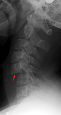
1. Complete anterior dislocation of affected vertebral body by half or more of the vertebral body AP diameter.
2. Disruption of the posterior ligament complex and the anterior longitudinal ligament.
3. "Bow tie" or " bat wing" appearance of the locked facets.


Question 9: Clay Shoveler's fracture is caused by:
Hyperflexion.
Hyperextension.
Axial compression.
Lateral rotation.
Explanation: Clay Shoveler's is fracture of a spinous process C6-T1 caused by powerful hyperflexion, usually combined with contraction of paraspinous muscles pulling on spinous processes (e.g. shoveling).


Question 10: Jefferson fracture is treated by:
Halo immobilization for 12 weeks.
Halo immobilization for 6 weeks.
Philadelphia collar for 6 weeks.
Immediate surgical fusion.

Explanation: Jefferson's fracture is treated with halo immobilization for 12 weeks, which usually results in primary union of the ring of C1 and stability of C1 with respect to C2. Surgical fusion may be needed if there is atlantoaxial instability after removal of halo.