Chest Radiology > Anatomy > Mass Location
Mass
Location
![]()
Intraparenchymal vs. pleural vs. extrapleural
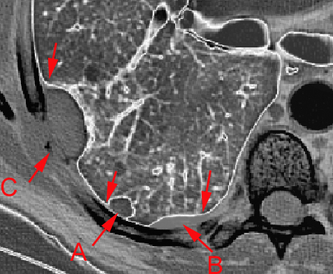
This diagram shows three locations that a mass can exist in the thoracic cavity.
A = intraparenchymal |
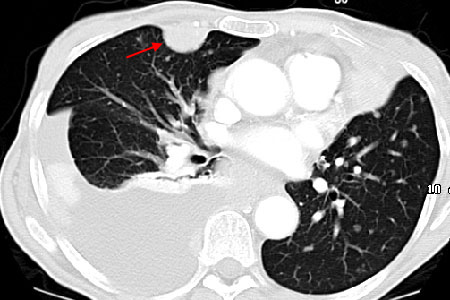
CT showing a mass that is likely pleural based (red arrow). Note the pleural effusion posteriorly.
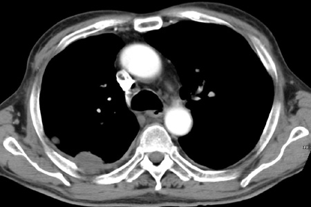
Another CT showing bone destruction indicative of an extrapleural mass.
![]()
![]()