Chest Radiology > Anatomy > Vasculature
Pulmonary Vasculature
![]()
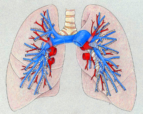
The following drawings show the major pulmonary vessels within the mediastinum. The bronchi that you have already learned are the same as on the prior drawing. These structures are obviously present on every chest x-ray but are usually unrecognized. If you learn the location of these structures, this will help you understand the anatomy as shown on chest x-rays and chest CT.

A drawing representing the pulmonary vasculature.
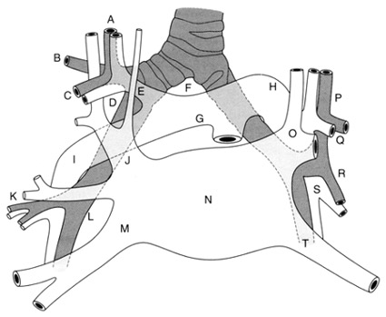
The following schematic drawing should help you sort out these structures. After the bronchi, remember that the left pulmonary artery arches over the left upper lobe bronchus and the right pulmonary artery passes posterior to the ascending aorta to divide into the truncus anterior and the descending RPA. Note that except in the right upper lobe, the pulmonary veins are generally anterior to the pulmonary arteries.
 |
A = Apical segmental
bronchus |


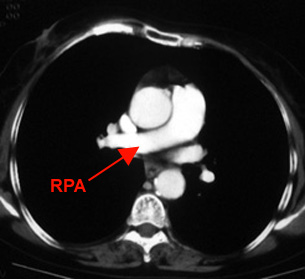
Left, left pulmonary artery on CT. Right, right pulmonary artery on CT. Note how the left pulmonary artery passes over the left mainstem bronchus to descend behind it, while the RPA passes behind the ascending aorta.
