Chest Radiology > Post-Test
Post-Test
![]()
1) Opacification of what part of the lung will silhouette the left heart border?
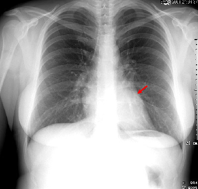
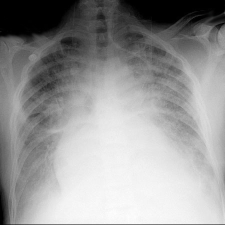
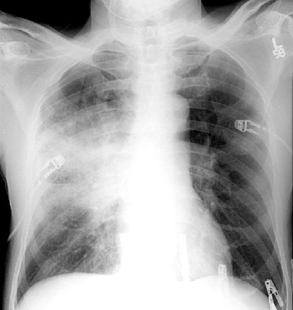
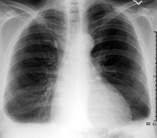
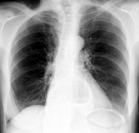
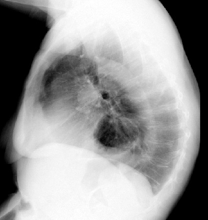
2) What is the most likely diagnosis on the following chest x-ray?
Questions 3-7: Please refer to the following image.
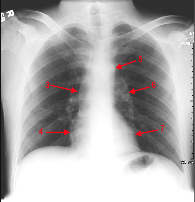
3) Identify the object labeled "3" in the above image.
4) Identify the object labeled "4" in the above image.
5) Identify the object labeled "5" in the above image.
6) Identify the object labeled "6" in the above image.
7) Identify the object labeled "7" in the above image.
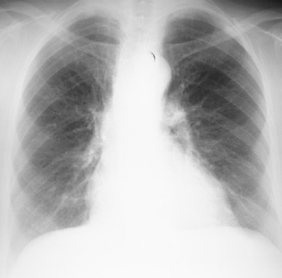
8) The normal chest x-ray seen below is not technically adequate. Why?
9) Which of the following is not a characteristic of a technically adequate PA chest radiograph?
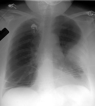
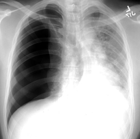
10) Identify the abnormality shown in the image below.
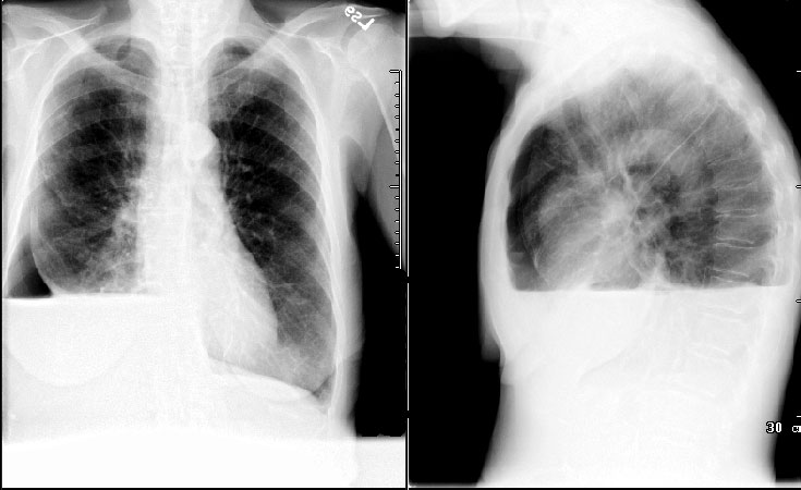
11) Admission PA and Lateral chest radiograph of a trauma patient with a left-sided pleural effusion demonstrates what other notable abnormality ?
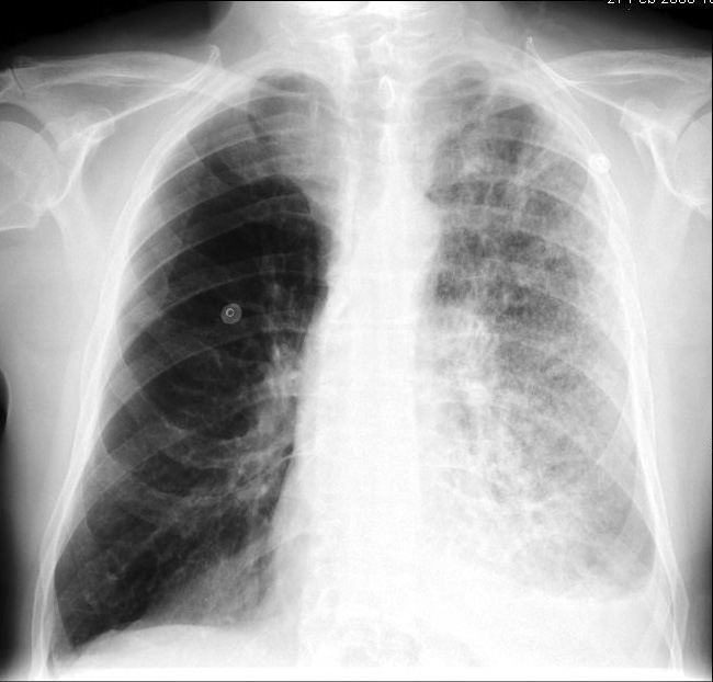
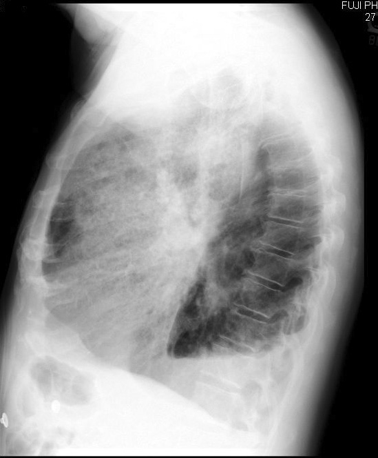
12) Based on your findings, what lobe is involved in the above image?
13) Name the abnormality shown in the image below?
14) What other clinical finding(s) would you expect to find in a patient with the abnormality shown above?
15) Identify the abnormality shown in the image below?
16) What study may be used to further evaluate the nature of the finding seen above?
17) Pneumonia causes volume loss or collapse of the affected lung parenchyma.
18) A pneumothorax is best demonstrated by an upright expiratory chest x-ray.
19) Identify the abnormality shown in the image below.
20) The next step in diagnosis and treatment of the abnormality seen above is?
21) Why is prompt diagnosis and treatment of the above process important?
22) The image seen below is a magnified view of a lateral chest x-ray. The blue arrows indicate the left ribs and the red arrows indicate the right ribs. Which of the following statements is true?

23) Identify the bronchus labeled "23" in the image below.
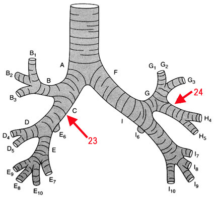
24) Identify the bronchus labeled "24" in the image above.
25) Identify the abnormality shown in the images below.
26) Which of the following statements is true when distinguishing between a pulmonary, pleural, and extrapleural mass?
27) What sign is seen in the image below?

28) Identify the abnormality shown in the images below.
29) The following characteristics of calcification in a solitary pulmonary nodule would be considered benign except:
30) What finding is least consistent with lobar atelectasis?