Emergency Ultrasound > Post Test
Post Test
![]()
All of the material covered in this test can be found on the previous pages. Answers are provided at the end of the test.
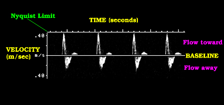
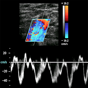
1) Which form of Doppler display is being demonstrated by the following image?
2) Which type of transducer gives the best overall image quality?
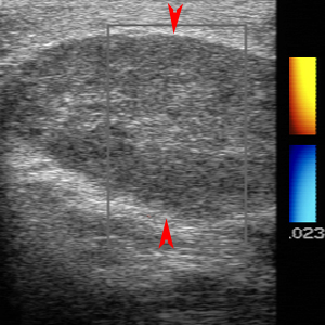
Questions 3-7: Please refer to the following image.
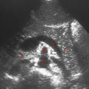
3) Identify the object labeled "3" in the above image.
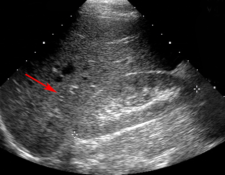
4) Identify the object labeled "4" in the above image.
5) Identify the object labeled "5" in the above image.
6) Identify the object labeled "6" in the above image.
7) Identify the object labeled "7" in the above image.
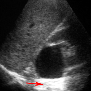
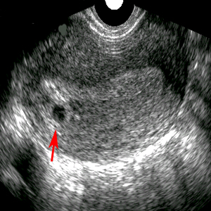
8) What is the red arrow indicating?
9) The best image resolution is obtained when using the highest frequency.
10) What is the optimal Doppler angle?
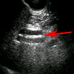
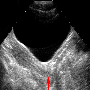
11) What is the red arrow demonstrating?
12) Which of the following is not part of the diagnostic criteria of acute cholecystitis?
13) What is labeled by the red arrow?
14) Which of the following is not a sonographic finding of acute pancreatitis?
15) If an object moves away from the transducer, the wavelength decreases and the frequency increases.
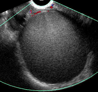
16) What is the red arrow indicating in the image?
17) Ultrasound is very reliable for demonstrating renal stones > 5 mm.
18) For the examination of the kidney, the best position to begin the exam is with the patient in the supine position.
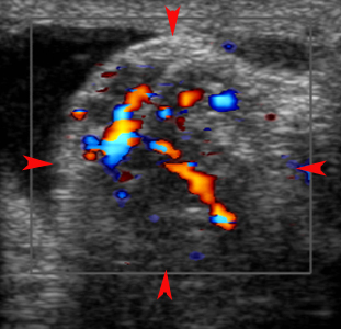
19) The image demonstrates appendicitis.
20) Which of the following is not a sonographic finding of a hemorrhagic cyst?
21) Hydronephrosis is a functional finding, not an anatomical finding.
22) A functional cyst is a smooth, round, anechoic, thin-walled structure larger than 2.5 cm.
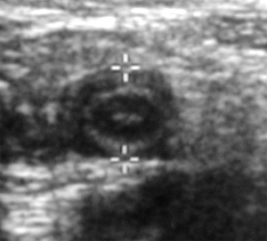
23) Identify the abnormality shown in the image below.
24) Fetal heart rate can be visualized by US around 6 weeks of gestation.
25) Identify the abnormality shown in the images below.
26) Deep venous thrombosis of the jugular vein is often asymptomatic.
27) A normal femoral vein in the adductor canal may not compress with transducer pressure.
28) Identify the abnormality shown in the images below.
29) The following image demonstrates normal flow in a peripheral vein.
30) Identify the abnormality in the following image.
![]()
![]()