Emergency Ultrasound > RUQ Pain > Acute Cholecystitis
RUQ Pain - Acute Cholecystitis
![]()
Clinical
Acute cholecystits is most commonly casued by impaction of a gallstone in the gallbladder (GB) neck obstructing the GB and resulting in inflammation of the GB wall. Ischemia and infection may result. Patients present with pain, right upper quadrant tenderness, and leukocytosis. About 70% of patients with acute cholecystitis have diffuse wall thickening. This finding, however, is neither sensitive nor specific for cholecystitis. Diffuse and marked wall thickening can also be seen in ascites, pancreatitis, hepatitis, CHF, sepsis, PUD and AIDS.
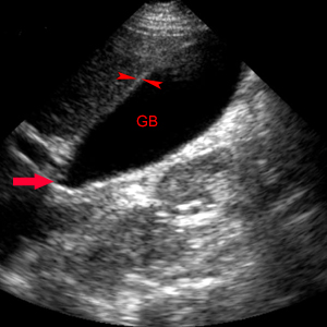
Image through the long axis of the GB (GB) demonstrates the gallbladder neck (red arrow). GB wall thickness is measured between the gallbladder lumen and the hepatic parenchyma (red arrowheads) with normal thickness < 3mm.
Exam
Begin the exam with the patient in the supine position. The patient can be moved to the left posterior oblique or upright position to demonstrate stone mobility. Image the full length of gallbladder from the portal vein to fundus with transverse images obtained at representative levels. Measure GB wall thickness perpendicular to wall adjacent to liver. Show the full length of CBD or as much as possible, and measure the CBD. Acquire longitudinal and transverse views of pancreatic head. Document any stones or biliary dilatation. If stones are seen, evaluate if they are mobile or impacted. Move the patient into upright or lateral decubitus positions to demonstrate stone mobility. Evaluate for focal and maximum tenderness over the gallbladder (Murphy's sign) by applying pressure with the transducer in the right costal margin.
Diagnosis for Acute Cholecystitis:
Major Criteria
Gallstones
Sonographic Murphy's sign
Minor Criteria
Wall thickening > 3 mm
Pericholecystic fluid

The gallbladder (GB) is filled with echogenic sludge (Sl) and a gallstone (red arrow) is impacted in the gallbladder neck. The gallbladder wall (red arrowheads) is markedly thickened indicative of wall edema and there are pericholecystic fluid (blue arrows) pockets surrounding the gallbladder.
![]()
![]()