Skeletal Trauma > Post Test Answers
Post Test Answers
![]()
1) Identify the fracture pattern shown in the image below.
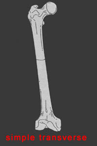
2) Identify the fracture alignment shown in the image below.

Questions 3-7: Please refer to the following image.
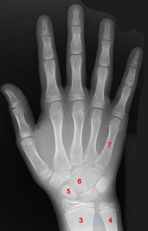
3) Identify the object labeled "3" in the above image. Radius
4) Identify the object labeled "4" in the above image. Ulna
5) Identify the object labeled "5" in the above image. Scaphoid
6) Identify the object labeled "6" in the above image. Capitate
7) Identify the object labeled "7" in the above image. 5th Metacarpal
8) Name the abnormality shown in the image below.
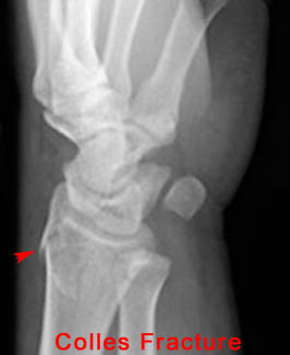
9) When evaluating the fracture shown in the question above, what secondary fracture should also be looked for?
Ulnar Styloid Process Fracture - In 50% of Colles fracture cases the ulnar styloid process is also fractured.
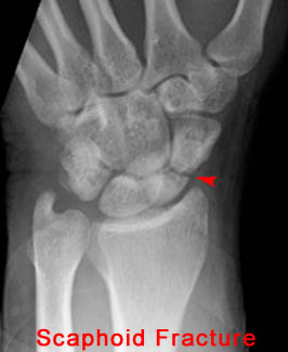
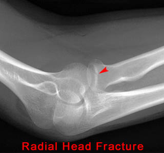
10) Name the abnormality shown in the image below.
11) What abnormalities of the elbow can be observed in the image below?
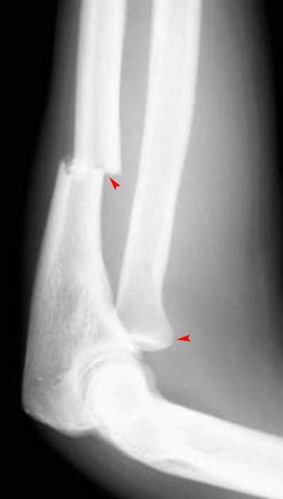
The arrows on the image above indicate an Ulnar Fracture and Radial Head Dislocation.
12) Based on your findings, what fracture is shown in the above image? Monteggia Fracture
13) Name the abnormality shown in the image below.
14) What other clinical finding(s) would you expect to find in a patient with the fracture shown above?
Soft Tissue Abnormalities
15) Name the abnormality shown in the image below.

16) What anatomical structure can be easily damaged during a dislocation such as the one shown above?
Neurovascular Bundle
17) Type I acromioclavicular joint separation is defined as a partial tear of the acromioclavicular ligament with no displacement. Under such conditions, x-ray of the region would be abnormal.
False - The first part of the statement is true. However, under such conditions x-ray of the region would be normal, not abnormal.
18) In cases of thoracic and lumbar spine compression fractures, CT is indicated for assessment of the spinal canal.
True - CT should always be performed for full assessment of the spinal canal in compression fracture cases because neurological deficit occurs in 65% of patients.
19) Name the abnormality shown in the image below.
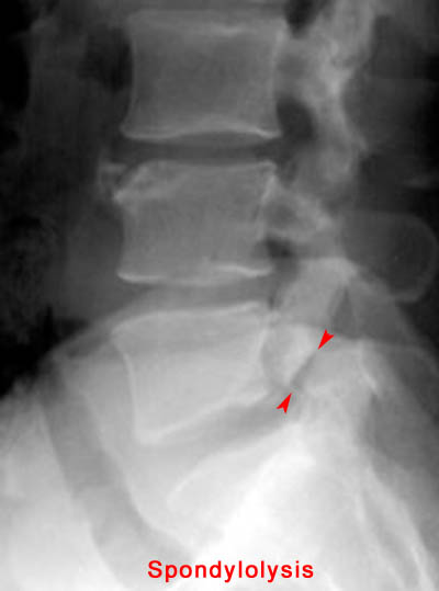
20) In which demographic is the injury shown above most common? Adolescent Athletes
21) At which spinal level is the injury shown above most common? L5
22) Name the abnormality shown in the image below.
Posterior hip dislocation.
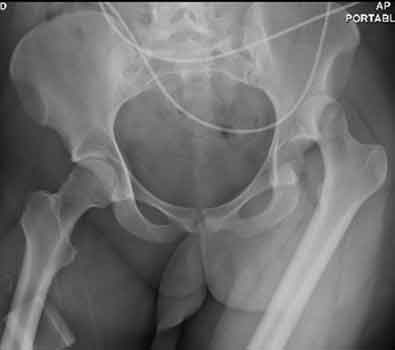
23) What abnormalities of the pelvis can be observed in the image below?
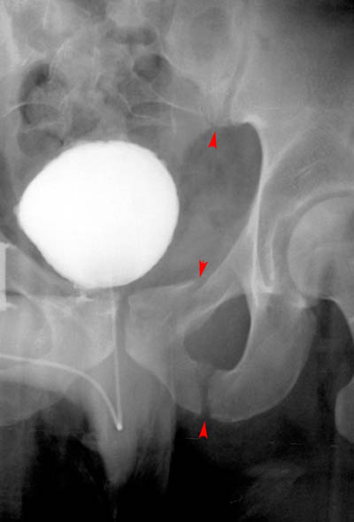
In the above image the sacroiliac joint and the ipsilateral ischiopubic rami are fractured, making the answer to the question "All of the Above."
24) Based on your findings, name the condition shown in the image above. Malgaigne Fracture
25) Name the abnormality shown in the image below.
26) How can a patellar fracture be distinguished from bipartite or multipartite patella?
Bipartite and multipartite patella have rounder edges that don't fit together as well.
27) A patient is known to have a torn ACL and presents for x-ray examination of the knee. Which of the following abnormalities is often associated with a torn ACL?
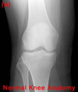
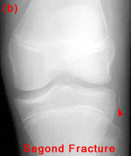
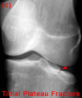
"B&C" - Both Segond fractures and tibial plateau fractures are commonly associated with ACL tears.
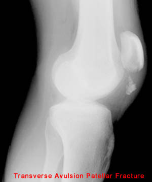
28) Name the abnormality shown in the image below.
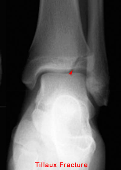
29) Give the specific name for the fracture type shown in the image above. Avulsion
30) A Jones Fracture specifically refers to a fracture of which metatarsal? 5th
![]()
![]()