- Pathogenesis:
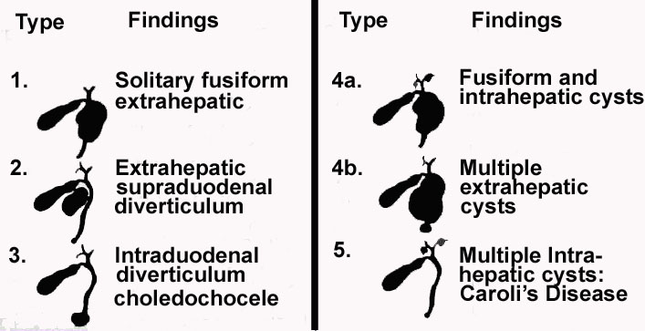
- There are five types of choledochal cysts. The fifth type, known as Caroli's disease, will be discussed on the following page.
- Choledochal cysts are the most common congenital bile duct anomaly and are often associated with other gallbladder anomalies, biliary stenosis, or atresia, and congenital hepatis fibrosis.
- 60% present before the age of 10.
- Appear more often in females (3:1).
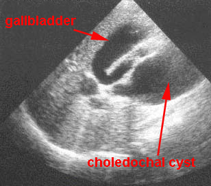
- Symptoms include obstructive jaundice in the neonate and right upper quadrant pain with intermittent jaundice and fever in older children and adults.
- Possible complications include the following:
- Rupture with secondary bile peritonitis.
- Cholangitis.
- Cirrhosis and portal hypertension.
- Calculus formation.
- Portal vein thrombosis.
- Liver abscess.
- Hemorrhage.
- Malignant transformation.
- Treatment involves surgical resection.
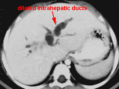
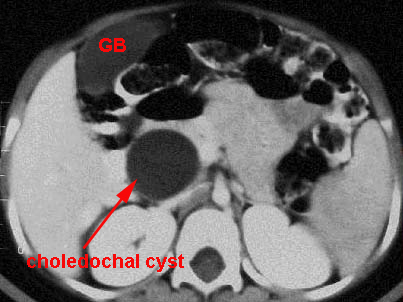
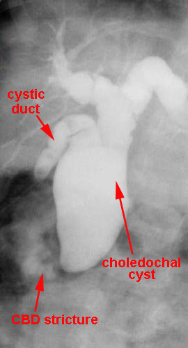
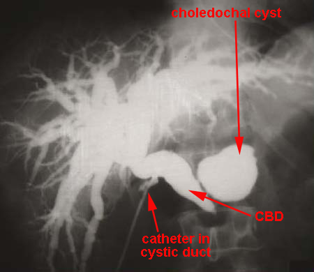
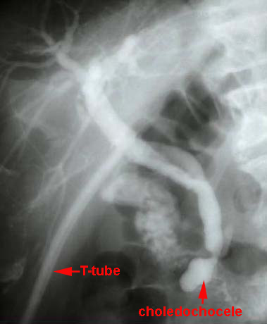
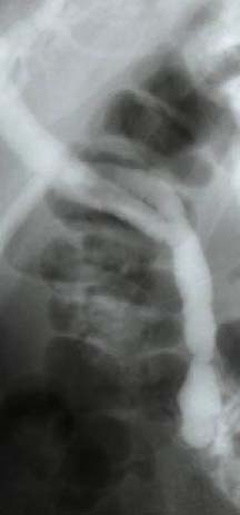
- Radiographic findings:
|