GI Radiology >
Biliary >
Gallbladder >
Neoplastic >
Porcelain Gallbladder
Neoplastic
Diseases of Gallbladder

Porcelain Gallbladder
|
- Pathogenesis:
- Unknown etiology, most likely due to chronic gallbladder inflammation and gallstones. Women are most frequently affected.
- Porcelain gallbladder refers to the calcification in the wall of the gallbladder. The metaphor refers to the brittle nature of the calcified wall.
- Usually, the patient does not have many symptoms, and the case is discovered upon incidental radiograph or CT.
- A patient with porcelain gallbladder almost always has gallstones along with cystic duct obstruction.
- Calcification may occur within the muscular layer or within the glands of the gallbladder mucosa.
- Calcification within the muscular layer is usually continuous and smooth.
- 10 to 20% of all patients with porcelain gallbladder develop gallbladder cancer during their lifetime—prophylactic cholecystectomy is recommended.
- Radiographic findings:
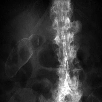
- Plain film: extensive calcification is apparent around the perimeter of the gallbladder.
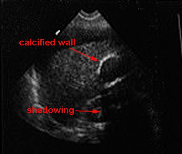
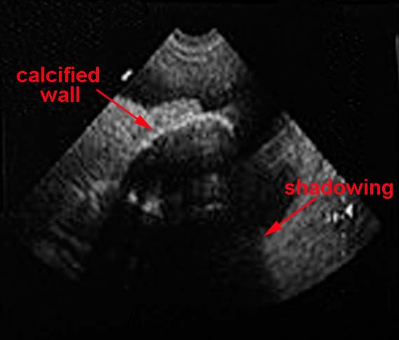
- U/S: bright, echogenic gallbladder wall with posterior shadowing in the gallbladder fossa.
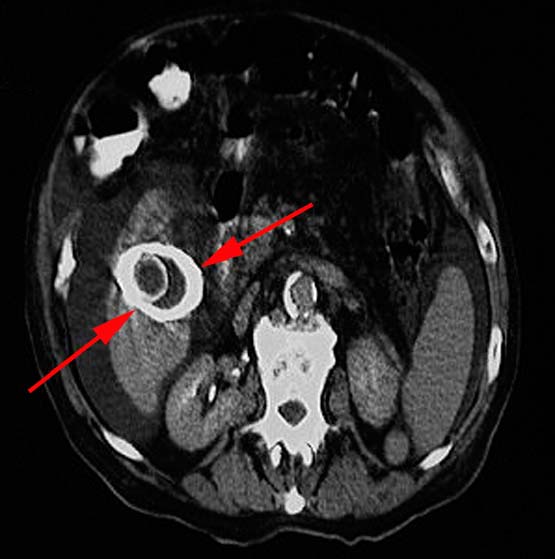
- CT: Calcification of the gallbladder wall (arrows), often accompanied by stones.
|
|

