GI Radiology > Biliary > Quiz Answers
Biliary Anatomy Quiz Answers
![]()
|
1. The upper limit of normal for the dimensions of a normal gallbladder are: A. 10 cm long, 3 cm wide B. 15 cm long, 5 cm wide C. 5 cm long, 3 cm wide D. 20 cm long, 5 cm wide E. 15 cm long, 10 cm wide A: The dimensions of a normal, non-dilated gallbladder are 10 cm long x 5 cm wide. Dimensions larger than these parameters are considered dilated, and could indicate underlying pathology or obstruction. |
|
2. (T/F) The gallbladder is divided into three fundamental regions: fundus, body, and neck. True False TRUE: These are the fundamental regions by which gallbladders are assessed. Review the gallbladder anatomy section. |
|
3. (T/F) Approximately 80% of stones are lucent cholesterol and 20% contain enough calcium to be detected on radiographs. True False TRUE: The majority of gallstone are undetectable on plain films due to the predominance of the lucent cholesterol stones. The imaging modality of choice for evaluation is therefore ultrasound. |
|
4. (T/F) Porcelain gallbladder has a gender bias, most frequently affecting males. True False FALSE: Considering all patients with porcelain gallbladders, there is slight female predominance. The etiology is unknown. |
|
5. Which of the following is the most likely long-term complication of porcelain gallbladder?
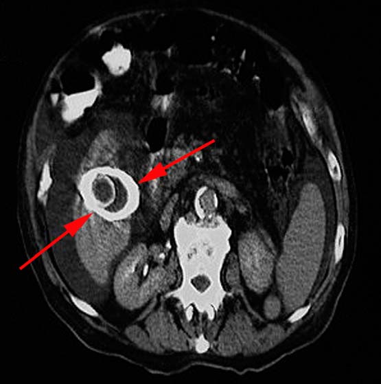
A. Gallbladder necrosis B. Biliary Atresia C. Gallbladder Cancer C: The image shown is an axial CT slice demonstrating the concentric calcification of the gallbladder wall, often accompanied by stones. 10 to 20% of all patients with porcelain gallbladder develop gallbladder cancer during their lifetime, therefore prophylactic cholecystectomy is recommended. |
|
6. What condition does the following image most likely represent? A. Ascending Cholangitis B. Biliary Obstruction C. Mirizzi's Syndrome
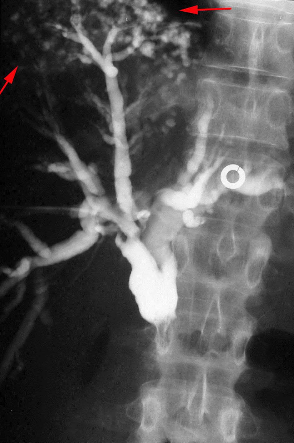
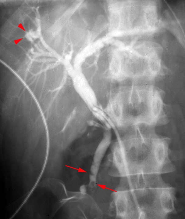
A: The pathogenesis of ascending cholangitis is gram (-) bacterial infection. Patients present with abdominal pain, fever, and obstructive LFTs. Associated radiographic findings early during the process reveal nothing but an obstructive pattern. Later findings reveal pleating or wall irregularity, mural necrosis and even intrahepatic abscesses at duct ends (as demonstrated by red arrows on the right image above). |
|
7. Which of the following predisposes to emphysematous cholecystitis? A. Diabetes B. Jaundice C. Hyperthyroidism D. Collagen Vascular Disease E. none of the above A: Emphysematous Cholecystitis is defined as gas in the gallbladder wall as a result of infection. The condition is a sequela of gallbladder ischemia with secondary infection by Clostridia, Escherichia coli, Staphylococcus, or Streptococcus, gas producing organisms. This condition is seen in patients with diabetes and is more common in men. |
|
8. (T/F) Patients with emphysematous cholangitis have an increased risk of perforation and treatment usually requires emergent cholecystectomy. True False TRUE: This condition is considered an emergency, requiring immediate cholecystectomy due to an increased risk of perforation. |
|
9. (T/F) Choledochal cysts are the most common congenital bile duct anomaly. True False TRUE: Choledochal cysts are the most common congenital bile duct anomaly. 60% present before the age of 10. Symptoms include obstructive jaundice in the neonate and right upper quadrant pain with intermittent jaundice and fever in older children and adults. |
|
10. (T/F) Patients with choledochal cysts have an increased incidence of other gallbladder anomalies, biliary stenosis/atresia, and congenital hepatic fibrosis. True False TRUE: Choledochal cysts are often associated with other gallbladder anomalies, biliary stenosis, or atresia, and congenital hepatis fibrosis. Additional possible complications include: rupture with secondary bile peritonitis, cholangitis, cirrhosis and portal hypertension, calculus formation, portal vein thrombosis, liver abscess, hemorrhage, malignant transformation. |
|
11. Caroli's Disease is a type of choledochal cyst characterized by: A. Solitary Fusiform Extrahepatic Cyst B. Extrahepatic Supraduodenal Diverticulum C. Intraduodenal Diverticulum Choledochocele D. Multiple Intrahepatic Cysts D:
|
|
12. Which of the following is NOT a predisposing condition for cholangiocarcinoma: A. Primary Sclerosing Cholangitis B. Choledochal Cyst C. Congenital Hepatic Fibrosis D. Clonorchis infection E. Exposure to Thorotrast F. Aflatoxin F: Aflatoxin exposure predisposes to hepatocellular carcinoma; not cholangiocarcinoma. All other choices increase risk of cholangiocarcinoma. |
|
13. (T/F) Choledochal cysts are more common in females. True False TRUE: Choledochal cysts demonstrate a female predominance (3:1). |
|
14. (T/F) Cholangiocarcinoma is the most common primary malignancy of the biliary system. True False TRUE: Cholangiocarcinoma is the most common. By the time lesions are found, they are often advanced, commonly extending along the duct and spreading to regional lymph nodes. |
|
15. (T/F) Cholangiocarcinomas are typically situated as extrahepatic lesions vs. intrahepatic, True False TRUE: Cholangiocarcinoma is usually extrahepatic rather than intrahepatic with the extrahepatic distal common biliary duct being affected in 50%-70% of cases. Klatskin's tumor refers to cholangiocarcinoma that arises at the bifurcation of the left and right hepatic ducts. |
|
|


