GI Radiology > Colon > Congenital Anomalies
Congenital Anomalies
![]()
Malrotation |
|
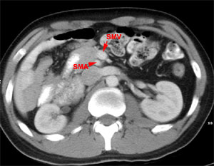
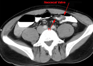
Malrotation occurs if the midgut does not complete the 270-degree rotation prior to re-entering the abdominal cavity during weeks 10-12 of gestation. The right colon and cecum overlie the duodenum in the right upper quadrant and Ladd's bands form over the organs to peritonealize the cecum. Infants may present with a flat abdomen, bloody stools, and bilious vomiting indicative of volvulus, ischemia, or proximal bowel obstruction. Malrotation is associated with intussusception, Hirschsprung's, intestinal atresia, and abdominal wall defects. Radiographically, patients will display signs of gastric or duodenal obstruction on plain films. An upper GI barium study will show displacement of the ligament of Treitz on the right, and a barium enema will show the cecum in the right upper quadrant. Treatment consists of surgery to correct the malrotation may involve severing the Ladd's bands, removing the appendix, reducing any volvulus, and fixing the duodenum in the right upper quadrant and the cecum in the left upper quadrant.
CT showing transposition of the SMA and SMV CT showing the ileocecal valve on the left instead of right side |