GI Radiology > Colon > Structural Abnormalities > Diverticulosis
Structural Abnormalities
![]()
Diverticulosis |
|
|
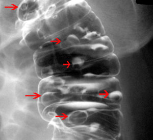
Diverticulosis is a condition in which the mucosa and muscularis mucosae herniate through the muscularis propria of the colon wall and produce a saccular outpouching. The major risk factor for developing diverticular disease is a low-fiber diet. The incidence increases with age, being rare before the age of 40 and affecting 50% of people over age 75. Only 10-15% of patients will develop symptoms of the disease like pain, diarrhea, melena, or distention. Approximately 70% of diverticula occur in the sigmoid colon and 25% in the ascending colon. Most appear on the antimesenteric side of the colon, between taenia. Plain films and barium studies reveal diverticula as gas/barium-filled sacs parallel to the lumen of the colon. Most are 5-10mm in diameter, but may range from tiny spikes to 2cm. The muscular layer may appear thickened with a distorted luminal contour in CT. Patients are managed with fiber supplements and stool softeners.
Diverticula on plain film (arrows) |

