GI Radiology > Colon > Vascular > Diverticular Hemorrhage
Vascular Complications
![]()
Diverticular Hemorrhage |
|
|
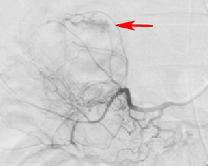
Diverticular Hemorrhage can develop from leakage of a vasa recta within a diverticula. 5% of patients with colonic diverticuli have massive bleeding and 45% have minor bleeding. Though most diverticula are the sigmoid region, approximately half of diverticular bleeds originate from the right colon. Patients will pass large volumes of bright red blood from the rectum, and will often be hypotensive and tachycardic. Syncope may occur. Colonoscopy is used to approach slow, intermittent hemorrhages, whereas angiography and scintigraphy are used to evaluate torrential bleeding. Angiography in conjunction with scintigraphy with 99mTc sulfur colloid or 99mTc-tagged erythrocytes confirms the presence of active bleeding and helps localize the site of hemorrhage. If the bleeding rate is greater than 0.5 ml per minute, selective mesenteric angiography may demonstrate extravasation of contrast agents. Management depends upon severity of the hemorrhage. A transfusion may be required depending on amount of blood loss. Persistent hemorrhage despite 2-4 units transfused blood is an indication for angiography and scintigraphy to localize the site of bleeding for semielective surgical resection of the involved colonic segment. In non-surgical candidates angiocatheters may be used to deliver intra-arterial vasopressin or synthetic emboli to stop the hemorrhaging. In extreme cases where the source cannot be determined, total abdominal colectomy may be necessary.
Inferior mesenteric angiogram demonstrating extravasation of blood during diverticular hemorrhage (arrow) |

