GI Radiology > Esophagus > Anatomy
Anatomy of the Esophagus
![]()
Normal Anatomy |
|
The esophagus is a muscular tube that is normally 25-30 cm long and 2-3 cm wide. The layers of the esophagus are the same as elsewhere in the gi tract with a mucosa, submucosa, circular and longitudinal layers of muscle, and adventitia. However, unlike the rest of the gi tract, the esophagus has no serosa. In the upper third of the esophagus the circular layer of muscle is made up of striated muscle while the lower two-thirds are made up of smooth muscle. The lining of the esophagus is made up of a thick mucosa that creates longitudinal folds that allow for distension. When relaxed, the mucosal surface is smooth. The esophagus is found anterior to the vertebral column and extends from approximately C6 down, following the curvature of the vertebral column. It passes through the esophageal hiatus of the diaphragm at approximately T10. The esophagus is divided into three segments: the cervical, thoracic, and abdominal segments. The cervical portion is separted from the cervical vertebrae by only a few mm of prevertebral soft tissue. It is also in contact with the trachea and the left mainstem bronchus, separated only by connective tissue. Spread of infectious and neoplastic processes are facilitated by the close relationship to these structures in addition to the fact that there is no serosa covering the esophagus. There are several structures that are in close proximity to the esophagus and which make normal impressions on it. These structures are the aortic arch at T3-T4, the left mainstem bronchus, the left inferior pulmonary veins and in some normal healthy people the left atrium, although a large impression is usually a sign of left atrial disease.
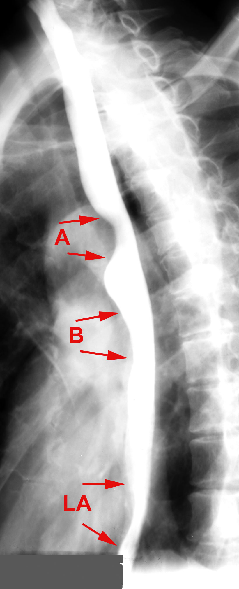
This oblique view of a normal barium swallow shows the normal impressions made by the (A) aortic arch, (B) left mainstem bronchus, and (LA) left atrium on the esophagus. |