GI Radiology > Esophagus > Congenital Esophageal Anomalies
Congenital Esophageal Anomalies
![]()
Esophageal Duplication |
|
Clinical Duplications can occur anywhere along the length of the gastrointestinal system. Theories as to how duplications occur include failure of recanalization, intrauterine vascular insults, and incomplete twinning. Duplications are located in continuity with or in close proximity to segments of the gut. A communicating duplication shares a wall or lumen with the adjacent "true" gastrointestinal tract. A noncommunicating duplication is adjacent to a segment of the "true" gastrointestinal tract, but does not share a lumen. The noncommunicating duplication are four times more common than the communicating variety. Noncommunicating duplications tend to be rounded and cystic. Most duplications are diagnosed before the age of 2. Clinically, a patient with esophageal duplication may present with respiratory symptoms of airway narrowing or it may be found incidentally on chest films. Radiographically, it is not possible to differentiate esophageal duplications from bronchiogenic cysts.
Radiological findings Prenatal ultrasounds may reveal fluid filled structures in the abdominal cavity. In the case of esophageal duplications, they may manifest as soft tissue masses on chest radiographs, suggestive of adenopathy or a posterior mediastinal tumor. If chest films demonstrate a mediastinal mass, a chest CT should be also obtained. The CT can show the density of the fluid in the cystic structure and also the extent of the duplication. Contrast studies may also be necessary in order to better assess any communication between the true esophageal lumen and the duplication.
The above PA and lateral views of the chest show a mediastinal mass.
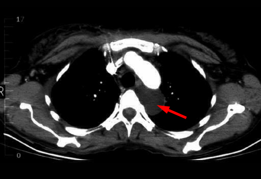
Follow
up CT of the chest shows an adjacent cystic structure that is found to
be a duplication cyst. |


