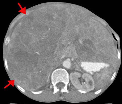
- Pathogenesis:
- Hepatic vein obstruction, usually
thrombosis, leading to portal hypertension and ascites.
- Key: large, tender liver with increased
ascites.
- Mostly idiopathic. Rare causes are
hypercoagulable states (e.g., polycythemia vera, malignancy, oral
contraceptive use) or local diseases (HCC, pancreatic cancer, or RCC).
- The caudate lobe is spared because its
venous drainage is directly to the IVC.
- Radiographic findings:
- Findings depend on the acuteness or
chronicity of the condition.
- Acute: dramatic decompensation; can go
into shock.
- Chronic: ascites and hepatomegaly.
Jaundice is less common.
- U/S: absent flow in the hepatic veins or
IVC; inhomogeneous echoes.
- CT: hepatosplenomegaly and patchy enhancement (arrows); the caudate lobe may also be enlarged in conditions longer than several weeks and collateral circulation may also be visualized.

- MRI: decrease in size and number of
hepatic veins and visualization of collateral circulation.
|
![]()
![]()