GI Radiology > Liver > Quiz Answers
Answers to Liver Quiz
|
1. The most cost-effective form of liver imaging for patients with RUQ pain and abnormal LFTs is: A. CT B. Ultrasound C. MRI B: Ultrasound is the first and most commonly obtained method for examination in patients with RUQ pain, abnormal LFTs, or suspected liver masses. It can evaluate bile duct obstruction and in combination with Doppler it may be used to evaluate vascular abnormalities as well. |
|
2. During what contrast phase is a hepatocellular carcinoma most likely to enhance? A. Arterial phase B. Portal venous phase C. Equilibrium phase A: Primary malignancies of the liver typically have hepatic arterial supply, thus will enhance during the arterial phase. Normal liver parenchyma has primarily portal venous supply, therefore, enhances during portal venous phase of I.V. contrast. |
|
3. The liver normally appears hyperdense to (brighter than) the spleen on CT. True False TRUE: The liver is normally more dense than the spleen. If it appears less dense than the spleen it is likely due to fatty infiltration. |
|
4. 75% of the blood suply to the liver is from the hepatic arteries. True False FALSE: 25% of the blood supply to the liver is from the hepatic arteries and 75% is from the portal vein. |
|
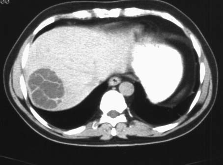
5. The following image depicts:
A. Focal nodular hyperplasia B. Hepatocellular carcinoma C. Simple liver cyst D. Echinococcal cyst D: This is an echinococcal cyst. Notice the calcified walls, membrane septation and focal areas of increased attenuation within the cysts that are typical. The cysts can rupture into the pleural cavity, peritoneal cavity, alimentary canal, or biliary tree, causing profound shock, peritonitis, and anaphylaxis. |
|
6. All of the following are signs of advanced cirrhosis on imaging EXCEPT: A. Liver surface nodularity B. Contracted liver with ascites C. Atrophy of the posterior segments (VI,VII) of the right lobe D. Enlarged caudate lobe (I) and lateral segments (II,III) of the left lobe E. Prominent umbilical vein F. Irregular enhancement G. All of the above are signs of advanced cirrhosis G: All of the above are signs of advanced cirrhosis on imaging |
|
7. The most sensitive and specific imaging modality for detecting focal fatty infiltration is: A. Ultrasound B. MRI C. CT B: MRI can use "chemical shift" or "in-phase and out-of-phase"gradient echo imaging to detect focal fat variation. |
|
8. The most common cause of a hyperechoic liver mass on U/S is: A. Hepatoma B. Simple cyst C. Hemangioma D. Echinococcal cyst C: Hemangiomas, benign proliferation of vascular tissue, are the most common cause of a hyperechoic liver mass on U/S. |
|
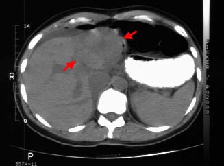
9. The following image depicts hepatic hemangioma.
True False FALSE: The image shows focal nodular hyperplasia. Noncontrast CT shows the lesion has low attenuation (arrows) compared with normal liver. The central stellate scar also shows low attenuation. |
|
10. All of the following are characteristic of hepatocellular adenomas EXCEPT: A. Benign lesions, but may be pre-malignant B. Risk of severe hemorrhaging C. Show marked enhancement on contrast CT D. Associated with oral contraceptives and steroids E. All of the above are associated with hepatocellular adenomas C: Typically, hepatocellular adenomas do not show marked enhancement on contrast CT, although hemorrhage may be seen. They typically have a heterogeneous and variable appearance secondary to fat and hemorrhage. |
|
11. Risk factors for developing hepatocellular carcinoma include: A. Cirrhosis B. Hemochromatosis C. Steroid use D. Hepatitis B or C E. All of the above E: Hepatocellular carcinoma typically occurs in an abnormal liver. All of the above cause changes to the liver that increase the risk of developing hepatocellular carcinomal. |
|
12. The appearance of the liver below is due to:
A. CHF B. Constrictive pericarditis C. Restrictive cardiomyopathy D. Any of the above D: The image depicts passive hepatic congestion, also known as nutmeg liver due to its appearance. The liver is enlarged without focal disease. The areas of low attenuation are caused by chronic increased hepatic venous pressure. |
|
|


