GI Radiology > Peritoneum > Infectious > Pseudomyxoma
Pseudomyxoma Peritonei
![]()
|
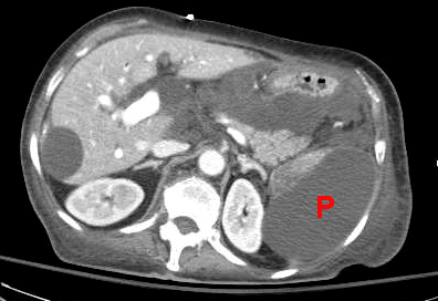
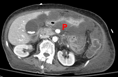
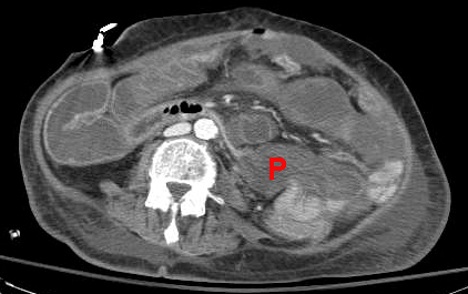
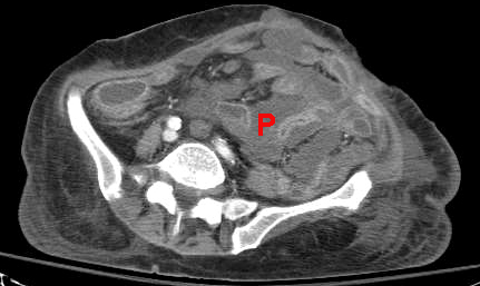
Clinical Pseudomyxoma peritonei is the filling of the peritoneal cavity with mucinous material from metastatic cystadenocarcinoma of the appendix of ovary. The morbidity and mortality from this process is very high.
Radiological findings The mucinous material forms gelatinous masses that appear cystic on CT and may calcify. The mucinous material will appear as high attenuation ascites along with thickening of the peritoneum. CT will also often show mottled densities, septations, and calcifications in the fluid. Mass effect on the liver and bowel is also often seen. Plain films may show punctate or ring-like calicifications scattered throughout the peritoneum. Ultrasound will show nodules that range from hypoechoic to strongly echogenic.
These CT images are from a patient with pseudomyxoma peritoneii. Notice how the abdomen is filled with the gelatinous material (P). It is difficult to distinguish the gelatinous material from the bowel in this patient without oral contrast. |