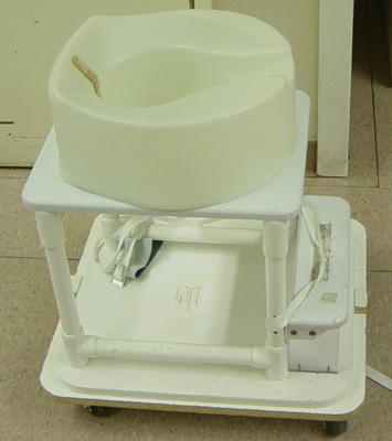
- Special commode.
- The patient is examined
in the lateral position while sitting on a commode which is specifically
designed for evacuation studies. The sitting position is more
physiologic than either the recumbent or standing position.
- The
specially constructed commode we use is the Brunswick Chair (E-Z-EM,
Inc., Westbury, NY). It has a radiolucent plastic seat that is filled
with water to provide increased soft-tissue density below the patient's
pelvis and buttocks. This filtration prevents burn-out of the image,
which occurs when there is too great a difference in radiolucency
between the high density pelvis and the external air. The seat has a
large central aperture into which a disposable plastic bag is fixed. The
commode has an adjustable footrest to recreate the patient's usual
position for bowel evacuation. For an examination, the commode is
mounted on the footrest of an upright fluoroscopy table.

- Disposable plastic seat cover/commode
liner for easy clean-up.
- Contrast medium.
- Accurate reproduction
of the physiologic conditions of defecation requires the use of a
contrast material with the consistency of normal stools - that is,
semisolid and malleable.
- Experiments have shown that defecography with
liquid barium underestimates the severity of abnormalities present, or
may even fail to demonstrate them, because fluid, like diarrhea, escapes
under the force of gravity or is evacuated without significant muscular
contraction.
- We use Anatrast (Lafayette Pharmaceuticals, Inc.,
Lafayette, IN) which is a barium sulfate paste (100% w/v) formulated to
simulate the consistency of stool. It is supplied in a 16 oz. tube. An
alternative choice is Evacu-Paste (E-Z-EM, Inc.), a barium sulfate paste
of 72% w/v.
- Administration system. The barium paste
is administered with a standard caulking gun which is attached to a
disposable enema tip.
- Recording Equipment.
- Static images are
obtained of the soft structures and bony landmarks while the patient is
(1) at rest, (2) during squeezing (voluntary contraction) of the pelvic
muscles, and (3) during evacuation.
- These images are obtained
using digital spot imaging.
- In addition, dynamic
recording of the evacuation phase of the fluoroscopic study should be
performed with a video recorder.
|
![]()
![]()