Inguinal Herniography (cont.)

Interpretation
|
|
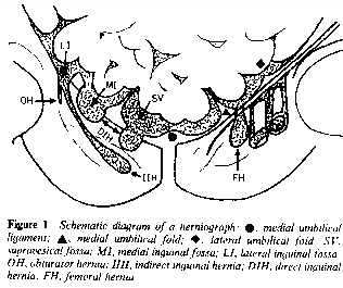
The inguinal herniogram is satisfactory
when the peritoneum is well outlined by contrast material both medial and
lateral to the notch formed by the inferior epigastric artery and vein
(Fig. 1, from ref. 4).

Indirect inguinal hernia.
- The vast majority of inguinal hernias are indirect.
- The basic defect is a patent processus vaginalis.
- This is seen as a peritoneal extension that begins lateral to
the inferior epigastric notch and passes obliquely downward and medial
into the inguinal canal. Depending on the extent of patency, it may
continue inferiorly and anteriorly to enter the scrotum.
- Strictly speaking, a hernia exists only when bowel or other
intraabdominal contents are present in the peritoneal sac.
Communicating hydrocele. A communicating hydrocele is a
potential hernia sac which results from the persisting processus vaginalis
and is filled with fluid.
Noncommunicating hydrocele is indicated when an
irreducible bulge is present clinically but is unfilled by contrast
material.
Direct inguinal hernia.
- Direct inguinal hernias are very uncommon.
- They result from a weakness in the transveralis fascia and protrude
directly through the external inguinal ring and are not related to a
persisting processus vaginalis.
- They are represented radiographically by a protrusion of contrast
which originates medial to the epigastric notch.
Femoral hernia.
- Femoral hernias are uncommon.
- They difficult or impossible to differentiate from the indirect
inguinal type.
- Whereas indirect inguinal hernias occur anterior and superior to the
inguinal ligament, femoral hernias occur posteriorly and inferiorly.
- Also, the patent processus vaginalis is usually tubular, whereas the
protrusion of peritoneum into the femoral canal is usually globular.
Obturator hernia. |
|
|


![]()

