GI Radiology > Small Bowel > Quiz > Case 2 Answers
Quiz: Case 2 Answers
![]()
5. With what imaging modality is this image acquired?
X-Ray
Fluoroscopy
CT
Nuclear medicine
Ultrasound
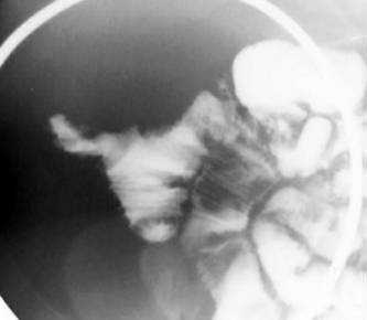
This spot film was taken during a single-contrast small bowel follow-through. Fluoroscopy offers much less spatial resolution than film-screen radiograph and exposes the patient to much higher doses of radiation. However, it allows for real-time evaluation (excellent for evaluating motility) and magnified images.
Note that the radiopaque circular object at the periphery of the image is the outline of the compression paddle.
6. What is the salient finding?
Dilated loops of bowel
Stricture
Fold thickening
Filling defect
Diverticulum
There is a finger-like outpouching emanating from a loop of small bowel, consistent with a diverticulum (Specifically, a Meckel's diverticulum). The other entities that can cause contrast material to reside outside the confines of the bowel lumen are ulcers, contained perforations, and fistulas. However, none of these would have this appearance. Note that the bowel caliber and fold thickness are normal.
7. What is most common sequela of a Meckel's diverticulum?
Asymptomatic
GI bleeding
Obstruction
Perforation
Cancer
Meckel's diverticulum is a relatively common congenital abnormality, reportedly present in up to 2% of the general population. This remnant of the vitelline duct invariably resides on the antimesenteric side of the distal ileum within two feet of the ileocecal valve. It most commonly presents as an incidental finding on imaging studies performed for other reasons. Symptomatic children usually present with obstruction, while symptomatic adults usually present with GI bleeding.
8. With what imaging modality is this image acquired?
X-Ray
Fluoroscopy
CT
Nuclear medicine
Ultrasound
This is an example of a nuclear medicine study. This image represents a Technicium-99m-tagged red blood cell scan ("Meckel's scan"). It demonstrates a focus of abnormal radiotracer uptake in the pelvis, which represents the Meckel's diverticulum. Note the relatively poor resolution but superb contrast of this image, resulting from selective uptake of radiotracer by the diverticulum.
9. What causes this test to be positive?
Infection
Ectopic gastric mucosa
Ectopic pancreatic mucosa
Carcinoid
Technetium-99m selectively localizes to gastric mucosa, salivary glands, and the thyroid. A large percentage of Meckel's diverticula contain ectopic gastric mucosa, which would cause a "positive" result on a Meckel's scan. Because bleeding from a Meckel's diverticulum is associated with ectopic gastric mucosa, this test is especially effective at evaluating for a Meckel's as the cause for GI bleeding. Remember also that GI bleeding is the most common symptomatic presentation in adults.
![]()
![]()