GI Radiology > Small Bowel > Quiz > Case 4 Answers
Quiz: Case 4 Answers
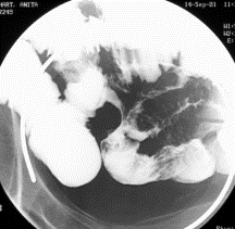
![]()
13. What is the salient abnormality?
Perforation
Diffuse fold thickening
Stricture
Filling defect
"Jejunalization"
The salient finding on this image is diffuse, regular fold thickening involving numerous loops of small bowel. This fold thickening is associated with luminal narrowing and separation of bowel loops.
This view is suboptimal for evaluating for free air. Perforation could very well be present but is not seen on this image. While strictures also cause luminal narrowing, they usually result in a featureless, "tube-like" appearance. No filling defects are seen. Jejunalization refers to a change in the appearance of the ileum, where it become more "feathery," while the jejunum conversely becomes more featureless. This finding is more typical of malabsorption syndromes, such as celiac or tropical sprue, and is not seen in this case.
14. What could cause this abnormality?
Mesenteric ischemia
Metastatic melanoma
Lympoid hyperplasia
Zollinger-Ellison Syndrome
Diffuse, smooth fold thickening is only caused by two things, edema and hemmorhage. Hypoproteinemic states and cardiac failure, as well as diffuse infectious, inflammatory (Crohn’s disease), or ischemic processes can result in diffuse bowel wall edema. An extreme form of bowel wall edema, shock bowel, occurs as a sequela of severe hypovolemia, as may be seen in severe trauma. Infectious, inflammatory, and ischemic processes are also implicated in diffuse bowel wall hemorrhage, as is hemorrhagic diasthesis and anticoagulant therapy. Radiation exposure (in large doses) can also cause both.
As the name implies, mesenteric ischemia results from inadequate blood supply to a portion of bowel because of occlusion or narrowing of a mesenteric artery. This case demonstrates occlusion of the superior mesenteric artery, which supplies the jejunum and ileum (in addition to the distal duodenum and proximal colon). Remember that ischemia can be acute or chronic. Acute mesenteric ischemia, which entails an acute vascular occlusion, gives rise to the clinical scenario of the acute onset of “abdominal pain out of proportion to the physical exam.” Chronic ischemia, however, is more indolent, occurring as a result of severe mesenteric arterial narrowing (usually from atherosclerosis). It presents clinically as postprandial abdominal pain (intestinal angina) and weight loss from fear of eating.
Metastatic melamoma presents as filling defects, variable in number and size. They can present as “target lesions,” signifying ulceration into the mass. Remember that a “target lesion” is highly suggestive of malignancy.
Nodular lymphoid hyperplasia presents as numerous tiny (2-4 mm) nodules. Note the well-described association between lymphoid hyperplasia, hypogammaglobulimemia, and giardiasis.
Zollinger-Ellison syndrome, caused by a gastinoma, usually presents with multiple ulcers and/or ulcers in atypical locations (e.g. postbulbar). Associated fold thickening is often present, but it is usually focal.
![]()
![]()