GI Radiology > Small Bowel > Congenital Anomalies > Annular Pancreas
Congenital Anomalies
![]()
Annular Pancreas |
|||
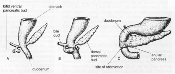
ClinicalAnnular pancreas is a rare
congenital abnormality in which a ring of pancreatic tissue encircles the
duodenum at or above the major papilla. Embryologically it is a sequelae of a
persistent left ventral bud, which usually atrophies during embryological
development.
The usual presentation of
annular pancreas is SBO. Annular pancreas compressing the second (descending)
portion of the duodenum. The degree of SBO caused by annular pancreas varies
dramatically from being asymptomatic to presenting as a surgical abdomen in a
newborn. It may become symptomatic in adulthood as SBO, with abdominal pain,
early satiety, and vomiting. There is also an increased incidence of peptic ulcer
disease and pancreatitis. Annular pancreas is discussed in more detail in the
Pancreas section. RadiologyPlain films may demonstrate
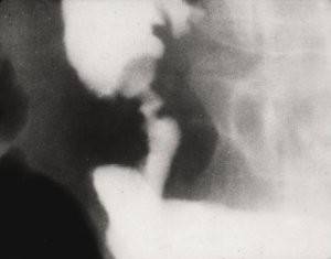
proximal small bowel obstruction. Fluoroscopy more clearly
delineates the abnormality. It will show dilatation of the proximal duodenum,
with eccentric or concentric narrowing of descending duodenum. In the most
severe cases, mucosal effacement will be seen. Note that there is NO
ulceration or mucosal destruction, differentiating it from neoplastic or
inflammatory etiologies. CT is beneficial for
diagnosis confirmation, as it will demonstrate the ring of pancreatic tissue
surrounding and compressing the duodenum.
Annular pancreas. Fluoroscopy demonstrates concentric
narrowing of the second portion of the duodenum. |
|||
|
|
![]()
![]()