GI Radiology > Spleen > Congenital > Accessory
Accessory Spleen(s)
![]()
-
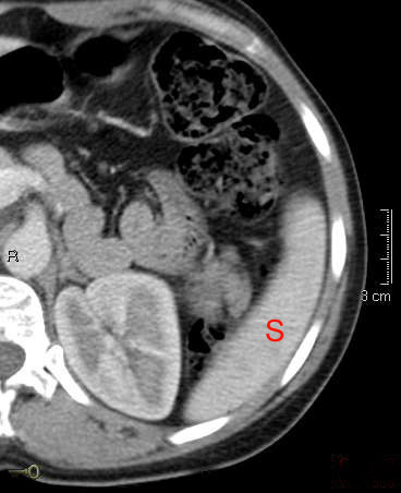
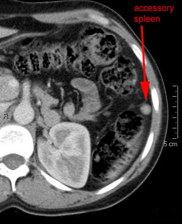
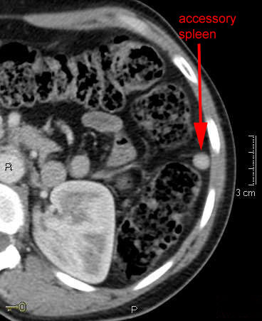
In embryological development, the spleen results from the coalescences of splenic buds. Failure of this process can lead to the formation of accessory spleens, typically seen in close proximity to the existing spleen. Accessory spleens can also be seen in other parts of the abdomen and retroperitoneum. These are incidental findings but these can be associated with recurrence of hematologic or pathologic disease after splenectomy, accessory spleen rupture, infarction, or torsion.


