Head CT > Infection > Subdural Empyema
Subdural Empyema
![]()
Subdural
empyema is usually due to meningitis, sinusitis, trauma or prior surgery.
It is a neurosurgical emergency. Subdural empyema leads to rapid clinical
deterioration, especially if it is due to sinusitis. On CT it appears
as an isodense or hypodense extra-axial mass. It has a lentiform or crescentic
shape.
The margin of collection often enhances with contrast material administration
due to the presence of granulation tissue or subjacent cortical inflammation.
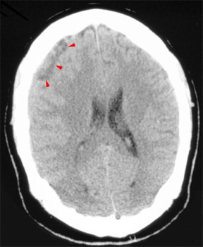 Notice the heterogeneous subdural fluid collection. 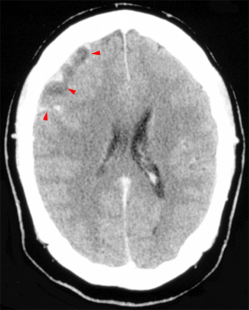
In the same patient,
post contrast administration, |
![]()
![]()