Head CT > Stroke > Subarachnoid Hemorrhage
Subarachnoid Hemorrhage
![]()
In
the absence of trauma, the most common cause of subarachnoid hemorrhage
is a ruptured cerebral aneurysm. Cerebral aneurysms tend to occur at branch
points of intracranial vessels and thus are frequently located around
the Circle of Willis. Common aneurysm locations include the anterior and
posterior communicating arteries, the middle cerebral artery bifurcation
and the tip of the basilar artery. Subarachnoid hemorrhage typically presents
as the "worst headache of life" for the patient. Detection of
a subarachnoid hemorrhage is crucial because the rehemorrhage rate of
ruptured aneurysms is high and rehemorrhage is often fatal.
CT is currently the imaging modality of choice because of its high sensitivity
for the detection of subarachnoid hemorrhage. CT is most sensitive for
acute subarachnoid hemorrhage. After a period of days to weeks CT becomes
much less sensitive as blood is resorbed from the CSF. If there is a strong
clinical indication, LP may be warranted despite a negative CT since small
bleeds can be unapparent on imaging.
On CT, a subarachnoid hemorrhage appears as high density within sulci
and cisterns. The insular regions and basilar cisterns should be carefully
scrutinized for subtle signs of subarachnoid hemorrhage. Subarachnoid
hemorrhage may have associated intraventricular hemorrhage and hydrocephalus.
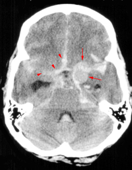 High density blood
fills the cisterns (arrowheads) in |
![]()
![]()