Head CT > Trauma > Subacute Subdural Hematoma
Subacute Subdural Hematoma
![]()
Subacute
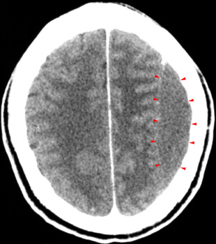
SDH may be difficult to visualize by CT because as the hemorrhage is reabsorbed
it becomes isodense to normal gray matter. A subacute SDH should be suspected
when you identify shift of midline structures without an obvious mass.
Giving contrast may help in difficult cases because the interface between
the hematoma and the adjacent brain usually becomes more obvious due to
enhancement of the dura and adjacent vascular structures. Some of the
notable characteristics of subacute SDH are:
- Compressed lateral ventricle
- Effaced sulci
- White matter "buckling"
- Thick cortical "mantle"

