Head CT > Trauma > Epidural Hematoma
Epidural Hematoma
![]()
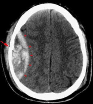
An epidural hematoma is usually associated with a skull fracture. It often occurs when an impact fractures the calvarium. The fractured bone lacerates a dural artery or a venous sinus. The blood from the ruptured vessel collects between the skull and dura. On CT, the hematoma forms a hyperdense biconvex mass. It is usually uniformly high density but may contain hypodense foci due to active bleeding. Since an epidural hematoma is extradural it can cross the dural reflections unlike a subdural hematoma. However an epidural hematoma usually does not cross suture lines where the dura tightly adheres to the adjacent skull.

