Pediatric Radiology > Chest > Pulmonary Inflammatory Disease > Tuberculosis


Pulmonary Inflammatory Disease

Specific Organisms


Tuberculosis remains a significant pulmonary infection in the 21st century, and the radiologist should maintain a high index of suspicion.

Primary TB in the pediatric population differs from the presentation of reactivation TB seen in adults. Primary TB produces a focal lobar consolidation in any pulmonary lobe. Hilar adenopathy and pleural effusions are common and should alert the radiologist to the possibility of TB.

If the lungs are secondarily infected hematogenously, miliary TB will result and present as characteristic, uniform small nodules diffusely through the lungs.


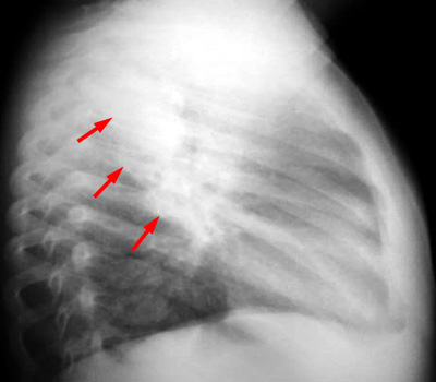
PA and LAT CXR with diffuse air space disease throughout the right upper lobe and significant right paratracheal adenopathy. The red arrow indicates adenopathy; the yellow arrow indicates TB pneumonia.