Pediatric Radiology > Genitorinary > Congenital Urinary Tract Anomalies
Congenital Urinary Tract Anomalies
![]()
|
Bilateral renal agenesis is present in 1 in 8000 births and is not compatible with a normal life. Unilateral renal agenesis is more common, present in 1 in 500 births. The ipsilateral adrenal gland is usually present and the contralateral kidney will demonstrate compensatory hypertrophy. If a normal kidney is not identified and renal agenesis is suspected a careful search will need to be made for a malpositioned kidney. Renal ectopia describes an abnormal position of the kidney. The kidney may migrate too far superiorly (thoracic kidney) or may not ascend completely (pelvic kidney). One or both kidneys may be on the contralateral side of the abdomen. This is seen with crossed fused ectopia where in most cases the kidneys are fused. The kidneys may also fuse across midline resulting in a horseshoe kidney. Horseshoe kidneys are more susceptible to trauma and are thought to be more prone to developing renal neoplasms. | |
 |
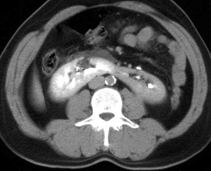 |
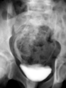
| Example of crossed fused ectopia on an IVP | Example of a horseshoe kidney on CT |
| Other congenital urinary tract anomalies include the following: | |
 |
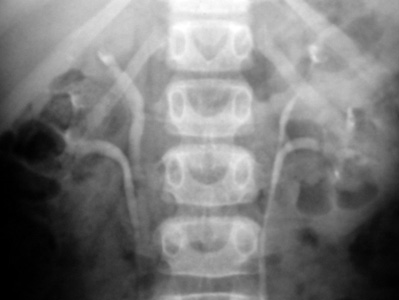 |
| Duplicate lower tracts | Duplicate upper tracts |
![]()
![]()