Pediatric Radiology > Genitorinary > Tumors > Imaging of Neuroblastoma
Imaging of Neuroblastoma
![]()
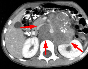
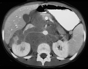
| Conventional radiographs will demonstrate an upper quadrant mass. Dystrophic calcifications will be present in 85% of cases. An excretory urogram will show displacement but not distortion of the collecting system. US, CT and MRI all have a role in imaging of neuroblastoma. Imaging is used to characterize the mass and define the extent of disease. The mass is most commonly suprarenal with displacement but no involvement of the kidney. Neuroblastoma will encase the vessels without any invasion or displacement. | |
 |
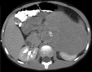 |
| Neuroblastoma with disease crossing midline | Neuroblastoma with calcifications |
 |
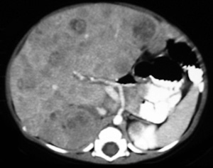 |
| Neuroblastoma encasing vessels | With liver metastasis |
![]()
![]()