Pediatric Radiology > Genitorinary > Tumors > Imaging of Wilms Tumor
Imaging of Wilms Tumor
![]()
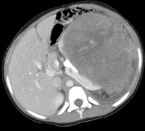
| Wilms tumor presents as a large mass with a mean diameter of 12 cm arising from the cortex of the kidney. Ultrasound is the first imaging modality used to evaluate an abdominal mass, although CT is frequently utilized. The "claw sign" will indicate the mass is arising from the kidney. In addition there will be intrinsic displacement of the renal collecting system. The mass will be hyperechoic on ultrasound with areas of heterogeneity. On CT Wilms tumor is a predominately solid enhancing renal mass. On both imaging studies there will be areas of heterogeneity due to hemorrhage and necrosis. Calcifications are uncommon, seen in less than 15%. On both imaging studies, it is important to look for invasion of the renal vein, IVC and right atrium. Pulmonary metastasis are relatively common (20%). | |
 |
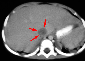 |
| Contrast enhanced CT of large mass arising from the kidney as evidenced by the claw sign and intrinsically displacing the renal collecting system. Note the absence of calcifications. | Contrast enhanced CT demonstrating tumor involvement of the IVC. |
![]()
![]()