Pediatric Radiology > Musculoskeletal > The Pediatric Hip > Slipped Capital Femoral Epiphysis
Slipped Capital Femoral Epiphysis
![]()
|
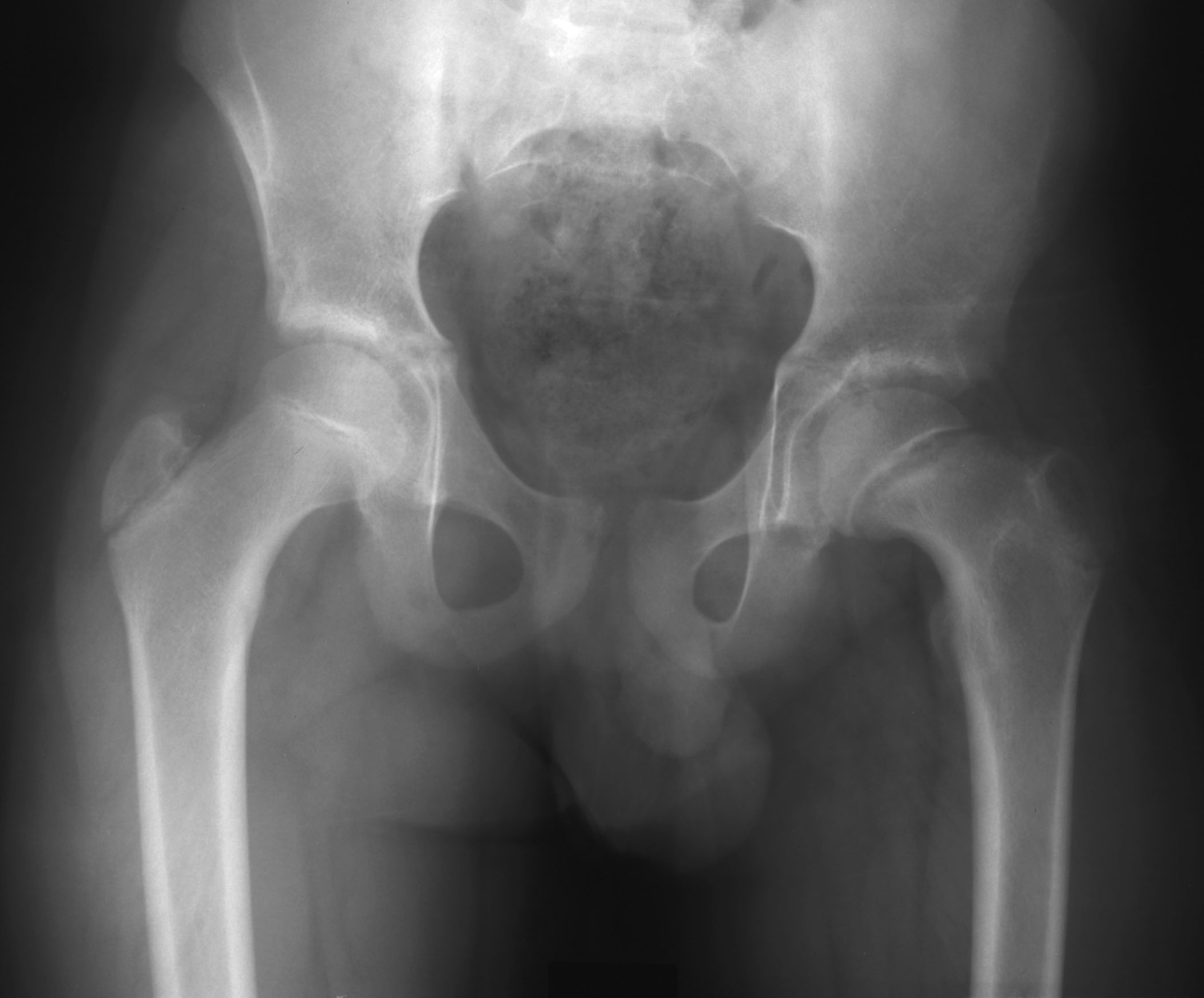
Slipped Capital Femoral Epiphysis (SCFE) is a Salter-Harris Type I fracture through the physeal plate of the proximal femur resulting in displacement. It should be suspected in any adolescent who complains of hip or knee pain. It is twice as common in males (12-15 years) than females (10-13 years). Predisposed risk factors include obesity, renal osteodystrophy, and endocrine disorders including hypothyroidism and hypopituitarism. Bilateral involvement of the hips can be seen in 20-30% of patients, but it is unusual to present at the same time. Displacement of the femoral head is posteromedial and often difficult to see on a standard AP film. Findings on AP film can include asymmetric physeal widening and/or an indistinct metaphyseal border at the level of the physis. Frogleg lateral views are often essential for diagnosis as minimal slippage can be better appreciated. Here, one can appreciate the posterior displacement of the epiphysis in relation to the metaphysis. On frogleg views, a line drawn tangential to the lateral cortex of the metaphysis should bisect a portion of the ossified epiphysis. If the epiphysis is medial to this tangential line, SCFE is the diagnosis. SCFE is typically treated surgically with pin fixation (done at current location) to prevent further slippage. Potential complications include avascular necrosis and chondrolysis.
|
||||

