Pediatric Radiology > Musculoskeletal > Metabolic Diseases > Sickle Cell Disease and Thalassemia
Sickle Cell Disease and Thalassemia
![]()
|
Sickle cell disease is a hemoglobinopathy in which polymerization of the prevalent HbS occurs, causing deoxygenated erythrocytes to lose their normal biconcave shape and become "sickle-shaped." Clinical manifestations of this disease include hemolytic anemia, infections, chest and abdominal pain, and skeletal pain (resulting from infarcts and osteomyelitis). Radiographic findings in sickle cell disease:
Thalassemia is a hereditary disorder resulting in decreased production of either alpha or beta globin chains. Radiographic findings in thalassemia:
|
|
|
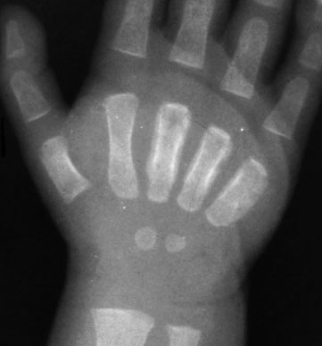
Sickle Cell "hand-foot syndrome" in an 8-month-old African American male with two week history of swollen hands and feet. AP radiograph of the right hand shows periostitis with subperiosteal new bone formation involving the 3rd-5th metacarpals. There are areas of lytic destruction (most noticeable involving the distal third metacarpal) possibly indicating an infarct. |
 |
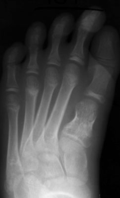 |
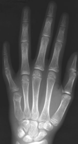
Chronic findings of "hand-foot syndrome" in a 12-year-old male with sickle cell disease. Left, Shortening of the 5th metacarpal of the left hand. Right, Shortening of the 1st metatarsal of the left foot. These findings are secondary to infarcts during bony growth. |
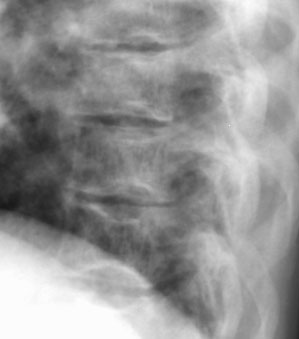 |
Sickle cell disease manifested in the spine of an adolescent female. Lateral thoracic spine film demonstrates "H-shaped" vertebral bodies. These findings represent compression infarcts of the end plates resulting from osteonecrosis of the vertebrae. |
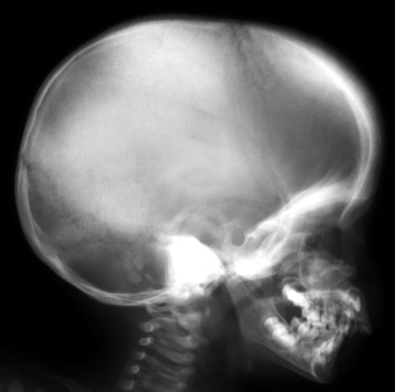 |
Thalassemia in a pre-adolescent male. Lateral skull radiograph shows a widened diploic space, especially in the frontal region. Also note the radiating spicules of bone extending at right angles to the inner-island tables, providing the "hair-on-end" appearance common in this and other forms of anemia. |
![]()
![]()