Pediatric Radiology > Neurological > Head Ultrasound
Head Ultrasound
![]()
Ultrasound is used to diagnose and follow complications of prematurity and screen for congenital abnormalities or hydrocephalus. The most common complications are germinal matrix hemorrhages and periventricular leukomalacia. Head ultrasound is performed in the neonate or infant through the open anterior fontanelle. Images are obtained in the sagittal and coronal planes. |
|
 |
 |
| Sagittal midline image | |
 |
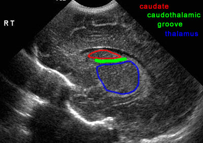 |
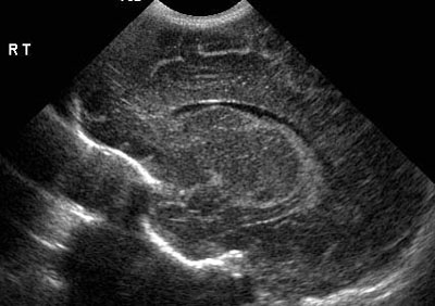
| Parasagittal image through caudothalamic groove | |
 |
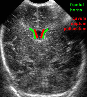 |
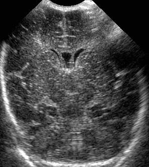
| Coronal image through frontal horns and cavum septum pellucidum | |
![]()
![]()