Pediatric Radiology > Neurological > Supratentorial Tumors > Parasellar > Craniopharyngioma
Craniopharyngioma
![]()
|
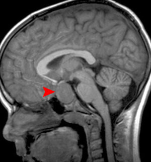
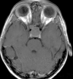
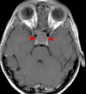
The most common parasellar mass lesion in childhood, craniopharyngiomas account for about 10% of childhood intracranial tumors. The most common presenting symptom is headache, often with nausea and vomiting. Half of children will have visual symptoms, most commonly bitemporal hemianopsia, decreased acuity, and double vision. They arise from persistence and proliferation of squamous epithelial cells within the tract of the embryologic structure, the craniopharyngeal duct. They may be entirely cystic or solid but most commonly are a combination of both. The cystic fluid may be high signal on all MRI sequences because of proteinaceous material. Eighty percent of cases demonstrate calcification on CT. Solid components typically enhance. |
|

|
|
| Sagittal MRI with contrast | |
|
|
| Axial MRI with contrast | |
|
Note: Sometimes difficult to differentiate from a craniopharyngioma, Rathke's cleft cyst can also occur in the same location and appear very similar on imaging and histology. Rathke's cleft cysts are remnants of Rathke's pouch between the pars distalis and the par nervosa of the pituitary. In contrast to craniopharyngioma, Rathke's cleft cysts do not calcify or enhance. Growth is unlikely and if present typically indicates a craniopharyngioma. |
|
![]()
![]()