ICU Chest Films > Lines and Tubes > Pacing Devices
Transvenous Pacing Devices
![]()
Patients in the ICU with bradyarrhythmias or heart block may require cardiac pacing. Transvenous pacers are introduced through the cephalic or subclavian vein into the apex of the right ventricle. Frontal and lateral projections are required to evaluate pacemaker placement. In the frontal view, the pacer tip should be at the apex with no sharp angulations throughout its length. On the lateral view, the tip should be imbedded within the cardiac trabeculae in such a way that it appears 3 to 4 mm beneath the epicardial fat stripe. A tip which appears beyond the epicardial fat stripe may have perforated the myocardium. Pacers placed within the coronary sinus will appear to be directed posteriorly on the lateral chest x-ray. The integrity of the pacer wire should be inspected along its entire length.
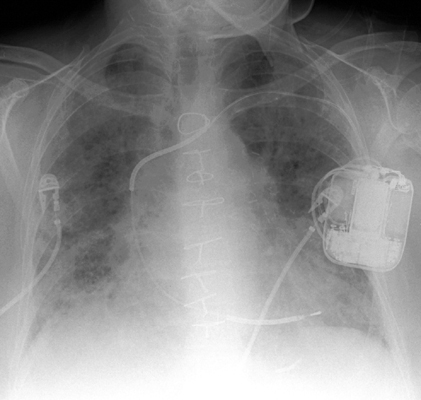
Single lead pacer with tip in the right ventricle.
![]()
![]()