Salter-Harris Fractures

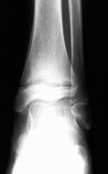
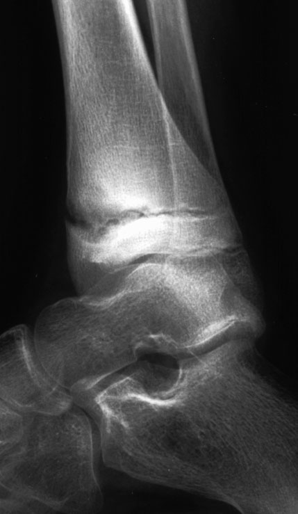
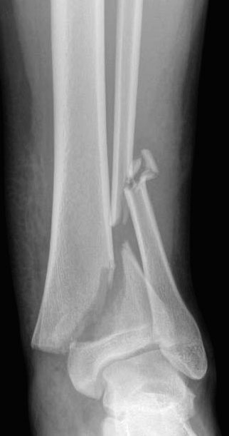
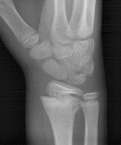
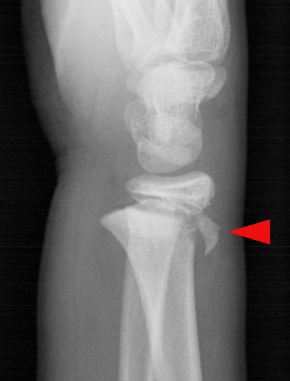
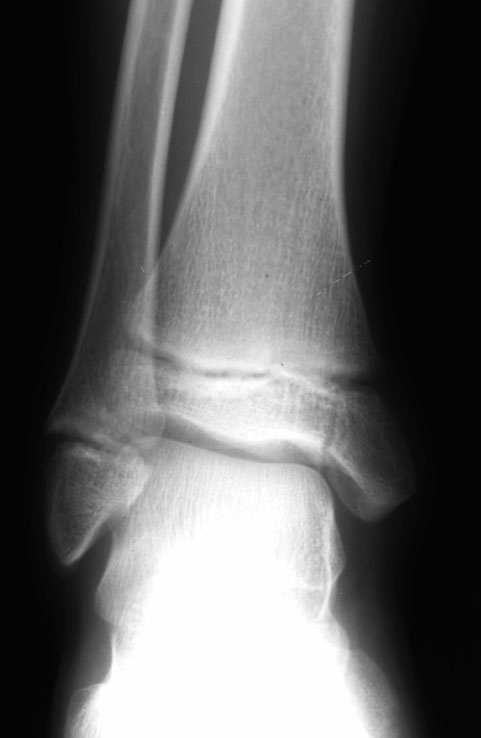
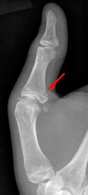
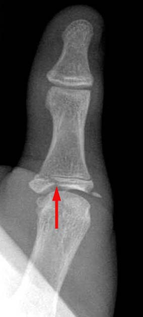
The
physeal plate is often involved in traumatic pediatric injuries. Up to
one-third of all pediatric fractures involving the long bones will involve
the physis. These fractures carry added significance because involvement
of the "growth plate" may lead to arrested development of the
affected limb. The more severe the injury, the higher the likelihood of
requiring surgery with internal fixation.
The standard
classification for physeal fractures was set forth by Salter and Harris.
This classification divides fractures into five types based on whether
the metaphysis, physis or epiphysis is involved as demonstrated radiographically.
|
Salter-Harris
Fracture Classification |
Description |
|
|
Type
I |
fracture
through the physeal plate (often not detected radiographically) |
Type
II |
fracture
through the metaphysis and physis (most common; up to 75% of all
physeal fractures) |
Type
III |
fracture
through the epiphysis and physis |
Type
IV |
fracture
through the metaphysis, physis and epiphysis |
Type
V |
crush
injury involving part or all of the physis |

 |
 |
Salter-Harris Type I fracture of tibial physis in a 10-year-old girl. AP
(left) and lateral (right) radiographs of right ankle demonstrate
a widened tibial physis. On the AP view, note the periosteal reaction
on the medial aspect of the distal tibial metaphysis. |
 |
Salter-Harris
Type II fracture of the left distal tibia.
AP radiograph of tibia shows a fracture-dislocation of the tibial physis
with a fracture through the metaphysis. Note the associated comminuted
fracture of the distal fibula. |
|
 |
 |
Salter-Harris
Type II Fracture of distal radius. Oblique (left) and lateral
(right) radiographs demonstrate a fracture of the radial physis with involvement
of the metaphysis. Note the fracture fragment (arrowhead) which is
displaced dorsally on the lateral image. |
 |
Salter-Harris
III fracture of distal tibial epiphysis in an 11-year-old boy. There
is a vertical fracture through the medial portion of the distal tibial
epiphysis. |
|
 |
 |
Salter-Harris IV fracture of the thumb. Two views of the left thumb reveal
a fracture at the base of the proximal phalanx (arrows), involving the metaphysis, physis, and epiphysis. |
|


© Copyright Rector and Visitors of the University of Virginia 2021