Cardiac MRI > Anatomy > Review Question Answers
Review Question Answers
![]()
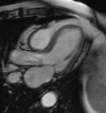
1) Which view is illustrated by the image below? Answer: A. This image demonstrates the three chamber view. It is obtained by selecting an image in the coronal plane through the aortic root and then choosing a plane which is perpendicular to the plane of the aortic valve. 2) Which of the following are obtained by using an image in the axial plane? Answer: C. The vertical long axis view is obtained from axial images. Additionally, the aortic view is also obtained from axial images. The next 3 questions refer to the following image: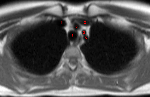
3) The left common carotid artery is labeled: Answer: C. The left common carotid is labeled "3" in the figure. 4) The label "5" in the figure is demonstrating which structure? Answer: C. The structure labeled "5" is the left subclavian artery. 5) True or false: The structure labeled "4" is a venous structure. Answer: A. True - the structure labeled "4" in the figure is the right brachiocephalic vein. |
![]()
![]()