Cardiac MRI > Anatomy > Vertical Long Axis View
Vertical Long Axis View
![]()
The vertical long axis is for evaluating the anterior and inferior walls and apex of the left ventricle. An axial image through the LV and LA is chosen from the transverse localizer images and a parasagittal plane that is perpendicular to the chosen image is prescribed that bisects the mitral valve and intersects the LV apex.
Mouse over the vertical long axis image to display labels.
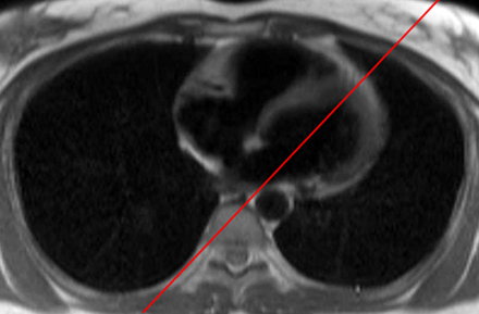

The red line in the figure on the left represents the plane through which the vertical long axis image on the right is obtained.
ANT = Anterior wall, AP = Apex, INF = Inferior wall, LA = Left Atrium, LV = Left Ventricle, MV = Mitral Valve
![]()
![]()