Cardiac MRI > Indications > Advantages & Disadvantages
Advantages and Disadvantages of Cardiac MRI
![]()
There are many advantages to Cardiac MRI when compared to other noninvasive imaging modalities such as ultrasound and CT. MRI does not have any ionizing radiation, thus permitting its use in children and pregnant women. It can produce high resolution and 3D images of the cardiac chambers and thoracic vessels. MRI gadolinium contrast media does not have as high a risk of allergic reaction or contrast media induced nephropathy as iodinated contrast media. Unlike echocardiography, MRI can produce images of cardiovascular structures without interference from adjacent bone or air which limits echocardiography. MRI is also less operator dependant than echocardiography. SSFP techniques can be used to assess global and regional ventricular contractile function, including the more difficult to assess right ventricle. Velocity encoded techniques permit measurement of blood flow. MRI does not have the weakness of geometric assumptions (as do angiography and 2-D echocardiography) in assessing ventricular volumes.
However, Cardiac MRI has several disadvantages. MRI requires more patient cooperation than other tests and claustrophobic patients may be unable to undergo the exam. Examination times are significantly longer as compared with CT. This, in addition to the fact that the patient is physically isolated from direct care when in the scanner, makes MRI unsuitable for unstable patients. Installation and operation of MRI equipment is costly and as such is an important consideration both for hospitals and patients. MRI is incompatible with various medical and life support devices, limiting its use in acute trauma. Due to the forceful attraction of ferromagnetic objects to the magnet, most intracranial or intraocular metal or shrapnel, cardiac pacemakers or pacemaker wires, and cochlear implants are absolute contraindications to imaging with MRI. Most new aneurysm clips, stents and vascular filters are MR compatible. Finally, MRI has less spatial resolution than CT, which limits the evaluation of small structures such as the coronary arteries.
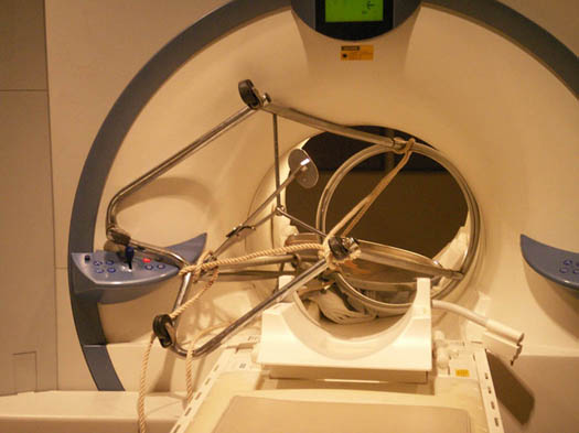
Ferromagnetic objects can be drawn into the magnet and cause injury to the patient. Likewise, any ferromagnetic material within the patient's body can be attracted to the magnet. Thus, an MRI imaging facility must diligently adhere to MR safety protocols.
![]()
![]()