Cardiac MRI > Pathology > Congenital Heart Disease > Anomalous Pulmonary Venous Return
Anomalous Pulmonary Venous Return
![]()
This congenital abnormality results in total (TAPVR) or partial (PAPVR) anomalous connection of the 4 pulmonary veins draining into the right heart circulation instead of the left atrium. The pulmonary veins can either enter the right atrium, SVC, or IVC below the diaphragm. Subdiaphragmatic APVR often causes vascular obstruction that leads to pulmonary edema in the lung drained by the anomalous pulmonary vein. This complex anatomy can be visualized effectively with cardiac MRI using cine imaging and MRA.
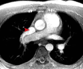
This gradient echo image in the axial plane shows the right superior pulmonary vein emptying into the SVC (arrowhead) rather than the left atrium. Therefore, oxygenated blood is entering the right side of the heart instead of the left. Other slices should be examined to study the course of the other pulmonary veins to determine if this is total or partial APVR.
![]()
![]()