Cardiac MRI > Pathology > Congenital Heart Disease > Acyanotic Congenital Heart Disease
Acyanotic Congenital Heart Disease
![]()
Acyanotic congenital heart lesions involve shunting of blood from the left heart to the right heart and most typically include atrial septal defect (ASD), ventricular septal defect (VSD), and patent ductus arteriosus (PDA). Acyanotic lesions always result in increased pulmonary blood flow. Cine gradient echo can be used to demonstrate turbulent flow from blood passing through a shunt. VENC imaging can be used to measure flow through the pulmonary arteries and aorta, thus allowing for quantification of the shunt fraction (Qp/Qs � pulmonic flow to systemic flow). 3D MRA imaging can help visualize the complex anatomy.
First pass of contrast agent through the heart. After the right atrium (RA) is filled, but before the left atrium (LA) is filled, unenhanced blood from the LA enters the RA via a atrial septal defect (ASD).
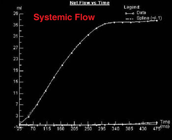
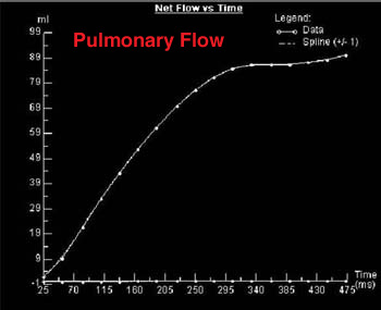
These Flow vs. Time graphs were obtained from the same patient using VENC imaging to determine the flow in the systemic and pulmonary systems. There is approximately twice as much flow in the pulmonary system (note the y-axis quantities), which yields a Qp/Qs ratio of about 2:1. Pulmonary flow greater than systemic flow is characteristic of a left to right shunt.
![]()
![]()