Cardiac MRI > Pathology > Congenital Heart Disease > Cyanotic Congenital Heart Disease
Cyanotic Congenital Heart Disease
![]()
Cyanotic congenital heart disease can occurs when blood from the right side of the heart enters the systemic circulation, resulting in cyanosis (SaO2 < 85%). The most common causes are Tetralogy of Fallot, Transposition of the Great Arteries, Total Anomalous Pulmonary Venous Return, Truncus arteriosus, Single ventiricle, and Tricuspid Atresia. Similar imaging techniques are used as in acyanotic congenital heart disease.
Ebstein's malformation occurs when there is congenital displacement of the septal and posterior leaflets of the tricuspid valve towards the RV apex. This results in tricuspid regurgitation and marked enlargement of the RA. Typically, there is an ASD present which allows for the right to left shunting of blood.

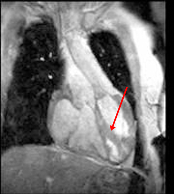
This gradient echo cine image in the coronal plane shows the great vessels arising in a parallel fashion, a finding that represents transposition of the great arteries (TGA). Notice that the ventricle on the patient's left is markedly trabeculated and a moderator band (arrow) is present; this therefore represents the morphologic right ventricle. In L-TGA , the morphological right ventricle is located in the usual location of the left ventricle and gives rise to the aorta. This configuration is also known a congenitally corrected transposition.
This gradient echo cine demonstrates some of the findings in Tetralogy of Fallot. The four findings in this condition are right ventricular hypertrophy, VSD, overriding aorta, and pulmonic stenosis. This cine shows all of these except the pulmonic stenosis since the pulmonary artery is out of the imaging plane.
