Cardiac MRI > Pathology > Cardiomyopathies > Hypertrophic Cardiomyopathy
Hypertrophic Cardiomyopathy
![]()
Hypertrophic cardiomyopathy (HCM) is a genetically acquired condition that results in hypertrophic myocardium. HCM results in diastolic heart failure due to impairment of myocardial relaxation during diastole. There are many different pheonotypes ranging from asymmetric to concentric hypertophy of the myocardium to just hypertrophy of a papillary muscle. The most common phenotype is asymmetric hypertrophy involving the interventricular septum. This asymmetric hypertophy may narrow the left ventricular outflow tract (LVOT), in which case it is referred to as hypertophic obstructive cardiomyopathy (HOCM).
Cardiac MRI is excellent at demonstrating the extent of hypertrophy in HCM, especially in the area of the ventricular apex. A flow void may be seen in the LVOT due to turbulent flow from narrowing, and VENC imaging can be used to quantify the increased velocity through the LVOT narrowing. Mitral regurgitation is frequently seen in patients with HCM. Systolic anterior motion of the mitral valve (SAM) is a phenomenon that occurs in the presence of LVOT obstruction, and is due to the anterior leaflet of the mitral valve getting sucked/pushed into the LVOT by accelerated flow. DHE should be performed on all patients with HCM due to the likelihood of scar tissue within the hypertrophied myocardium. This scar tissue can be a nidus for fatal arrhythmias. Cardiac MRI can be used to follow the patient after ventricular septal resection or percutaneous ablation.
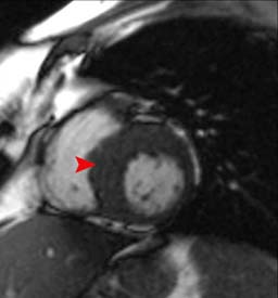
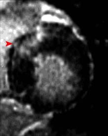
Asymmetric hypertophic cardiomyopathy involving the septum. The horizontal long axis (left) and short axis (middle) views demonstrate septal thickening. The lateral wall is normal in thickness. Delayed enhanced image (right) shows subepicardial and mid wall hyperenhancment of the antereoseptum representing fibosis.
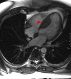
This three chamber view in a patient with HOCM shows a hypertrophied LV and abnormal systolic anterior motion (SAM) of the anterior mitral valve leaflet. It is this SAM that results in obstruction in the LVOT as demonstrated by turbulent flow (flow void).
![]()
![]()